Longitudinal Biomarker Assessment in Reproductive Aging: Discover Risks by Interpreting Multi-Hormone Patterns Over Time
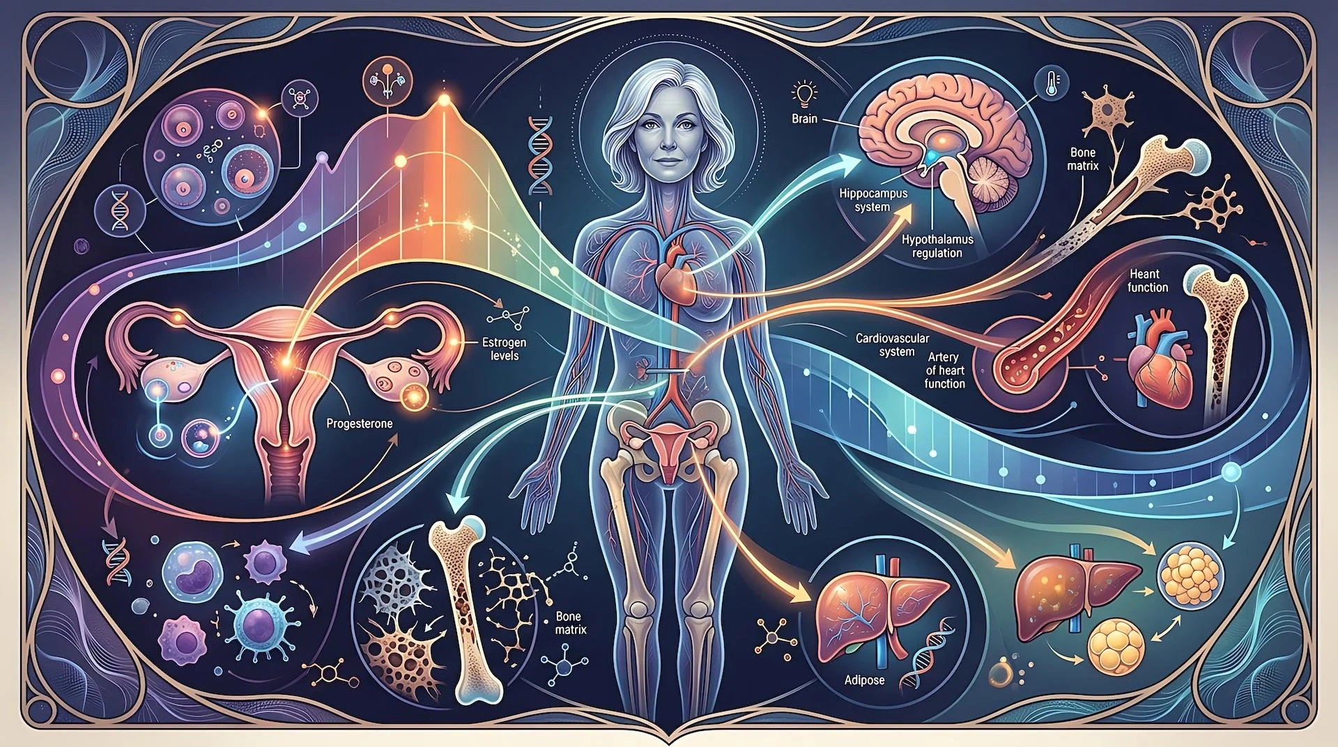
Perimenopause and menopause reflect progressive changes in ovarian function that alter communication between the ovaries, hypothalamus, and pituitary gland. These neuroendocrine adaptations begin years before the final menstrual period as ovarian reserve declines and follicular responsiveness changes.
Changes in estrogen, progesterone, follicle-stimulating hormone (FSH), and luteinizing hormone (LH) contribute to the hormonal variability of reproductive aging and influence menstrual patterns, sleep, mood, metabolism, bone remodeling, and vasomotor symptoms (1-3). While hormone testing provides valuable insight, individual values offer limited context; the clinical significance lies in recognizing patterns over time.
A pattern-based approach evaluates how these biomarkers change together across the menopause transition. E3G, LH, PdG, and FSH provide insight into ovarian function and physiological adaptation when interpreted longitudinally alongside symptoms and clinical history (3, 4).
The Hormonal Transition
Throughout reproductive years, the hypothalamic-pituitary-ovarian (HPO) axis coordinates follicular development, ovulation, and estrogen/progesterone production. As ovarian reserve declines, follicular responsiveness and ovulatory patterns become increasingly variable.
Key Hormonal Biomarkers
E3G (Estrone-3-Glucuronide) – E3G is a urinary estrogen metabolite that reflects ovarian estrogen activity and follicular recruitment. During early perimenopause, variability increases, with intermittent peaks due to irregular follicular activity. As ovarian function declines, estrogen activity decreases and stabilizes at a lower postmenopausal level.
Luteinizing Hormone (LH) – LH is a pituitary hormone that triggers ovulation. During the reproductive years, LH demonstrates a mid-cycle surge. During perimenopause, surges may become blunted or absent despite continued estrogen activity. Later stages are characterized by higher baseline LH without consistent ovulatory patterns (3).
Pregnanediol Glucuronide (PdG) – PdG is the primary urinary metabolite of progesterone and reflects ovulation and luteal function. A sustained post-ovulatory rise indicates progesterone production and luteal sufficiency. Declining PdG patterns reflect fewer ovulatory cycles and reduced progesterone exposure, which may contribute to changes in sleep, mood, bleeding patterns, and endometrial physiology (5).
Follicle-Stimulating Hormone (FSH) – FSH reflects pituitary signaling involved in follicular recruitment. As ovarian responsiveness declines and estrogen feedback decreases, FSH rises intermittently during early perimenopause before becoming persistently elevated after menopause. FSH is most informative when interpreted alongside E3G, LH, and PdG rather than independently (4, 6-8).
HORMONE PATTERNS ACROSS THE TRANSITION
Rather than following a predictable decline, reproductive hormones shift through recognizable patterns during the menopause transition. Interpreting E3G, LH, PdG, and FSH together provides a clinical framework for understanding changing ovarian function and the physiological adaptations associated with reproductive aging. Research demonstrates that the relationships among estrogen, progesterone, LH, and FSH evolve across transition stages, supporting a pattern-based approach to interpretation (3).
Early Perimenopause: Hormonal Variability
Early perimenopause is characterized less by declining hormone production than by increasing variability. During this time, estrogen activity often becomes more erratic, while progesterone production declines as ovulatory cycles become less frequent. In response to reduced ovarian signaling, FSH secretion increases to stimulate follicular recruitment, while LH patterns become increasingly variable (2).
E3G demonstrates intermittent peaks from irregular follicular recruitment, while ovulation becomes less consistent. LH surges may become variable, PdG rises shorter or lower, and FSH may show intermittent early-cycle elevations as pituitary drive increases.
This pattern may contribute to irregular bleeding, breast tenderness, migraines, mood fluctuations, and sleep disruption. Higher E3G with limited PdG support reflects estrogen exposure without consistent ovulation, potentially increasing endometrial stimulation when recurrent. Prospective studies have shown that declining estrogen together with rising FSH is associated with disrupted sleep continuity during perimenopause, even after accounting for vasomotor symptoms (5).
Fluctuating reproductive hormones may also influence immune signaling, inflammatory regulation, glucose metabolism, and stress physiology during the menopause transition (4, 6, 9).
Late Perimenopause: Increasing Anovulation
As the menopause transition progresses, anovulatory cycles become increasingly common. E3G generally trends toward a lower baseline, although intermittent estrogen surges may still occur. LH often demonstrates a higher baseline with fewer clearly defined ovulatory surges, PdG remains low across many cycles, and FSH becomes more consistently elevated.
This pattern reflects repeated anovulatory cycles in which intermittent estrogen exposure occurs without adequate progesterone opposition. In individuals with persistent irregular or prolonged bleeding, this pattern may warrant closer evaluation for endometrial health.
Declining estrogen signaling shifts bone remodeling toward greater resorption through increased osteoclast activity and altered bone turnover pathways (10). Reduced progesterone exposure may influence GABAergic signaling, contributing to sleep, mood, and stress-related changes, while declining estrogen contributes to vasomotor symptoms.
Higher FSH concentrations are associated with greater vasomotor, genitourinary, and psychological symptom burden during late perimenopause and postmenopause (11). Altered HPA-axis activity may further influence vasomotor symptoms, suggesting interaction between reproductive hormones and stress physiology (12).
Menopause: New Physiological Baseline
Following twelve consecutive months without menstruation, ovarian hormone production reaches a new physiological baseline. E3G stabilizes at a lower level, reflecting markedly reduced ovarian estrogen activity, while PdG remains persistently low because ovulation no longer occurs. LH remains elevated without cyclic surges, and FSH remains persistently elevated as pituitary signaling continues despite reduced ovarian responsiveness.
Although hormonal variability decreases after menopause, sustained reductions in estrogen and progesterone drive broader physiological adaptations, including increased bone resorption through osteoclast activity and altered skeletal pathways. Emerging research also suggests that immune signaling and the gut-bone axis may contribute to postmenopausal skeletal adaptation, with changes in microbial composition, intestinal immune regulation, and inflammatory pathways under investigation as contributors to bone loss (13-14).
Persistently lower estrogen activity also contributes to changes in vascular function, lipid metabolism, cardiometabolic health, and genitourinary tissues. In the central nervous system, reduced estrogen and progesterone signaling influences pathways involved in mood, cognition, and sleep, while reproductive aging has also been linked to changes in neuroimmune signaling and inflammation (9).
Patterns Requiring Closer Evaluation
While no single hormone defines reproductive aging, combinations of E3G, LH, PdG, and FSH, interpreted alongside symptoms and menstrual history, may reveal meaningful physiological patterns.
Persistently elevated FSH accompanied by lower E3G, persistently low PdG, and elevated LH is consistent with advancing ovarian aging. This pattern reflects diminished ovarian responsiveness and is commonly accompanied by increasing vasomotor symptoms, genitourinary changes, accelerated bone remodeling, and cardiometabolic adaptation. Higher FSH concentrations have also been associated with greater vasomotor, genitourinary, and psychological symptom burden during the menopause transition (11).
Intermittent high E3G peaks with absent or consistently low PdG suggest repeated estrogen exposure without consistent ovulation. This pattern reflects intermittent estrogen production without adequate luteal progesterone support and may occur as ovulation becomes increasingly inconsistent. Recurrent episodes may contribute to heavier or prolonged bleeding and increased endometrial stimulation, warranting closer evaluation when abnormal uterine bleeding is present.
Blunted or absent LH surges accompanied by persistently low PdG despite moderate E3G suggest repeated anovulatory cycles. These cycles are often associated with menstrual irregularity, cyclic migraine, mood fluctuations, and disrupted sleep as progesterone production becomes increasingly inconsistent.
When very low E3G, persistently elevated FSH and LH, and absent PdG occur before the expected age of menopause, broader evaluation may be appropriate to assess for premature ovarian insufficiency and other endocrine contributors.
Importantly, these hormone patterns are not diagnostic. Rather, they provide physiological context that should be interpreted alongside symptoms, menstrual history, age, and the broader clinical picture to guide clinical assessment and determine whether additional evaluation is warranted.
Clinical Context
Hormone patterns do not exist in isolation. The menopause transition reflects changing communication across multiple physiological systems, and factors beyond ovarian aging can influence both hormone dynamics and symptom expression. Interpreting E3G, LH, PdG, and FSH within the broader context of an individual’s health provides a more comprehensive understanding of reproductive aging.
Stress Physiology – Circadian rhythms and the hypothalamic-pituitary-adrenal (HPA) axis interact closely with reproductive hormone regulation. Altered cortisol rhythms, including reduced morning cortisol output and increased evening arousal, may amplify vasomotor symptoms, sleep disruption, and mood changes as progesterone and estrogen decline. Altered HPA-axis activity has also been associated with greater vasomotor symptom severity during the menopause transition (12).
Sleep Quality – Sleep is also shaped by the interaction of endocrine, psychological, and behavioral factors. Declining estrogen levels, depressive symptoms, anxiety, and stressful life events collectively influence sleep continuity and cognitive function throughout perimenopause (5, 15). Lower estradiol and higher FSH concentrations are associated with disrupted sleep continuity, highlighting the relationship between reproductive hormone dynamics and sleep physiology (5).
Thyroid Function – Thyroid dysfunction can produce symptoms that overlap with perimenopause, including fatigue, cognitive changes, menstrual irregularity, mood disturbances, and altered thermoregulation. Evaluating thyroid function alongside reproductive hormones helps distinguish overlapping endocrine patterns.
Gut-Liver Axis & Estrogen Metabolism – Following hepatic conjugation, estrogen metabolites enter the intestine, where microbial enzymes such as β-glucuronidase influence estrogen recycling. These microbial pathways, collectively referred to as the estrobolome, may contribute to variability in estrogen metabolism and symptom expression during reproductive aging (16-18).
Nutritional Status – Nutritional status also influences physiological adaptation during reproductive aging. Adequate intake of protein, magnesium, vitamin D, vitamin K, and zinc supports bone remodeling, neuromuscular function, and metabolic health as ovarian hormone production declines. Dietary patterns that promote microbial diversity may further support estrogen metabolism and immune regulation through emerging gut-endocrine interactions (18).
Clinical Interpretation
The clinical value of hormone assessment lies in understanding how E3G, LH, PdG, and FSH change together over time. These patterns provide context for menstrual changes, symptoms, and broader physiological adaptations throughout the menopause transition.
Patterns such as intermittent E3G elevations with limited PdG, persistently elevated FSH with declining E3G and PdG, or increasingly erratic LH activity can help characterize different stages of reproductive aging and identify physiological changes affecting endometrial health, sleep, bone remodeling, cardiometabolic function, and vasomotor symptoms. Integrating these trends with menstrual history, symptom presentation, and broader health factors supports a more individualized approach to menopause care.
Because reproductive aging is dynamic, longitudinal hormone assessment evaluates endocrine patterns rather than single measurements. A pattern-based approach recognizes menopause as a continuum involving sleep, metabolism, skeletal health, immune signaling, and neuroendocrine regulation.
The evolving approach will be explored in the upcoming clinical program: Advancing Hormone Assessment in Perimenopause and Menopause: Moving Beyond Symptoms to Longitudinal Monitoring
Join Dr. Carrie Jones, Dr. Tara Scott, and Dr. Michelle Leary during our free live webinar on September 8 from 5-7 PM Pacific Time for an evidence-informed discussion on advancing hormone assessment in perimenopause and menopause. This program will explore the interpretation of E3G, LH, PdG, and FSH patterns through longitudinal monitoring and how integrating hormone trends with symptoms and clinical history may support a more individualized approach to menopause care.
References
- Sherman BM, West JH, Korenman SG. The menopausal transition: analysis of LH, FSH, estradiol, and progesterone concentrations during menstrual cycles of older women. J Clin Endocrinol Metab. 1976 Apr;42(4):629-36. doi: 10.1210/jcem-42-4-629. PMID: 1262439.
- Weiss, G., Skurnick, J., Goldsmith, L., Santoro, N., & Park, S. J. (2004). Menopause and hypothalamic-pituitary sensitivity to estrogen.. JAMA, 292 24, 2991-6 . https://doi.org/10.1001/jama.292.24.2991
- Kawakita, T., Yasui, T., Yoshida, K., Matsui, S., & Iwasa, T. (2023). Associations of LH and FSH with reproductive hormones depending on each stage of the menopausal transition. BMC Women’s Health, 23. https://doi.org/10.1186/s12905-023-02438-5
- Wang, X., Zhang, H., Chen, Y., Du, Y., Jin, X., & Zhang, Z. (2020). Follicle stimulating hormone, its association with glucose and lipid metabolism during the menopausal transition. Journal of Obstetrics and Gynaecology Research, 46. https://doi.org/10.1111/jog.14297
- Coborn, J. E., De Wit, A. E., Crawford, S., Nathan, M., Rahman, S. A., Finkelstein, L., Wiley, A. S., & Joffe, H. (2022). Disruption of sleep continuity during the perimenopause: Associations with female reproductive hormone profiles.. The Journal of clinical endocrinology and metabolism. https://doi.org/10.1210/clinem/dgac447
- Zhang, C., Zhao, M., Li, Z., & Song, Y. (2020). Follicle-Stimulating Hormone Positively Associates with Metabolic Factors in Perimenopausal Women. International Journal of Endocrinology, 2020. https://doi.org/10.1155/2020/7024321
- Saini, P., Pathak, J., R., Goel, N., & Bhagat, D. (2025). Prevalence, severity, and association of serum follicle-stimulating hormone level with vasomotor and genitourinary symptoms in perimenopausal and postmenopausal women. Journal of Family Medicine and Primary Care, 14, 2934 – 2940. https://doi.org/10.4103/jfmpc.jfmpc_333_25
- Spicer, J., Malaspina, D., Blank, S. V., & Goosens, K. A. (2024). Follicle-stimulating hormone: More than a marker for menopause FSH as a frontier for women’s mental health. Psychiatry research, 345, 116239 – 116239. https://doi.org/10.1016/j.psychres.2024.116239
- McCarthy, M., & Raval, A. (2020). The peri-menopause in a woman’s life: a systemic inflammatory phase that enables later neurodegenerative disease. Journal of Neuroinflammation, 17. https://doi.org/10.1186/s12974-020-01998-9
- Xiong Q, Tang P, Gao Y, Zhang L, Ge W. Proteomic Analysis of Estrogen-Mediated Signal Transduction in Osteoclasts Formation. Biomed Res Int. 2015;2015:596789. doi: 10.1155/2015/596789. Epub 2015 May 18. PMID: 26120583; PMCID: PMC4450280.
- Saini, P., Pathak, J., R., Goel, N., & Bhagat, D. (2025). Prevalence, severity, and association of serum follicle-stimulating hormone level with vasomotor and genitourinary symptoms in perimenopausal and postmenopausal women. Journal of Family Medicine and Primary Care, 14, 2934 – 2940. https://doi.org/10.4103/jfmpc.jfmpc_333_25
- Sauer, T., Tottenham, L., Ethier, A. R., & Gordon, J. (2020). Perimenopausal vasomotor symptoms and the cortisol awakening response.. Menopause. https://doi.org/10.1097/gme.0000000000001588
- Tu Ye, Yang R, Xu X, Zhou X The microbiota-gut-bone axis and bone health. J Leukoc Biol. 2021; 110: 525–537. https://doi.org/10.1002/JLB.3MR0321-755R
- Ma Z, Liu Y, Shen W, Yang J, Wang T, Li Y, Ma J, Zhang X, Wang H. Osteoporosis in postmenopausal women is associated with disturbances in gut microbiota and migration of peripheral immune cells. BMC Musculoskelet Disord. 2024 Oct 7;25(1):791. doi: 10.1186/s12891-024-07904-1. PMID: 39375626; PMCID: PMC11460084.
- Chenji, S., Sander, B., Grummisch, J. A., & Gordon, J. L. (2024). Biopsychosocial factors intersecting with weekly sleep difficulties in the menopause transition.. Maturitas, 189, 108111 . https://doi.org/10.1016/j.maturitas.2024.108111
- Ervin SM, Li H, Lim L, Roberts LR, Liang X, Mani S, Redinbo MR. Gut microbial β-glucuronidases reactivate estrogens as components of the estrobolome that reactivate estrogens. J Biol Chem. 2019 Dec 6;294(49):18586-18599. doi: 10.1074/jbc.RA119.010950. Epub 2019 Oct 21. PMID: 31636122; PMCID: PMC6901331.
- Hu S, Ding Q, Zhang W, Kang M, Ma J, Zhao L. Gut microbial beta-glucuronidase: a vital regulator in female estrogen metabolism. Gut Microbes. 2023 Jan-Dec;15(1):2236749. doi: 10.1080/19490976.2023.2236749. PMID: 37559394; PMCID: PMC10416750.
- Wang H, Shi F, Zheng L, Zhou W, Mi B, Wu S, Feng X. Gut microbiota has the potential to improve health of menopausal women by regulating estrogen. Front Endocrinol (Lausanne). 2025 Jun 9;16:1562332. doi: 10.3389/fendo.2025.1562332. PMID: 40551890; PMCID: PMC12183514.