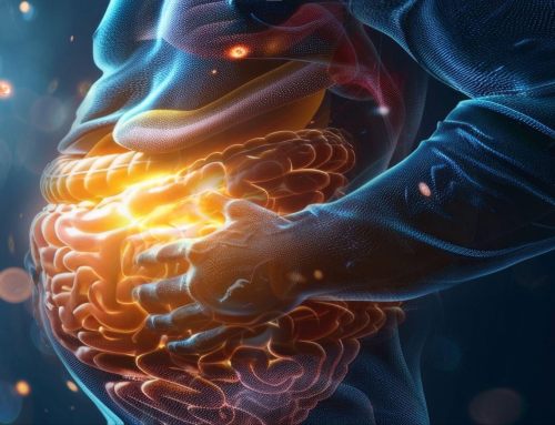
Immunomodulatory Interventions of Gut Mucosal Immune System Structures
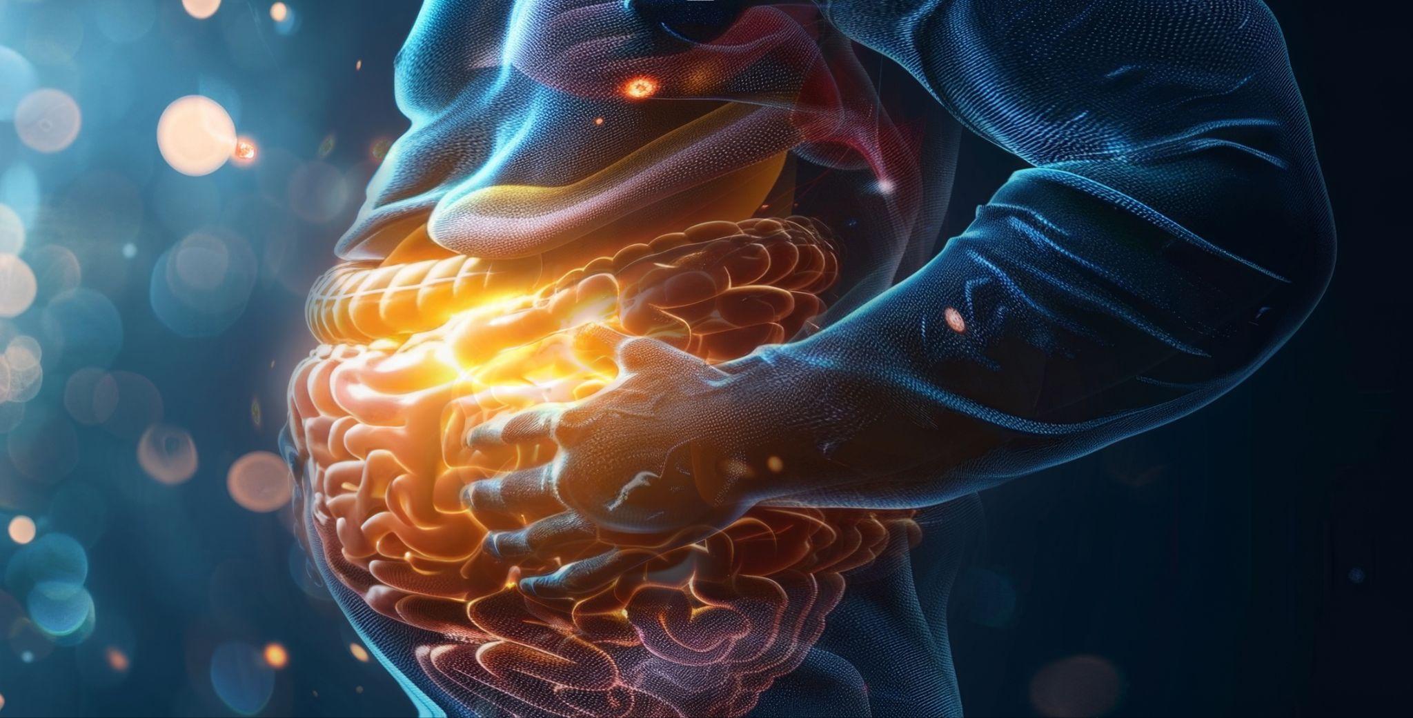
The mucosal immune system lies at points of entry to the body, where the inside meets the outside, such as the sinuses and the mouth. As the name implies, mucus is employed to defend the body at these points. The gut mucosal immune system (MIS) is one of the most complex points of entry. Tight control must be retained in the gut where food particles get absorbed, lest disease-causing agents sneak into the inner sanctum of the body along with life-giving nutrients. The immune system must allow passage of food particles but vigorously attack any disease, any pathogen. Unfortunately, the complexity of physiology and anatomy at these junctures increases the risk of something going awry.
Yet, the points of failure in any system are often where the opportunity for therapeutic intervention lies. Researchers have been relentlessly pursuing knowledge of the gut to address the growing numbers of gut-related illnesses that intersect with the immune system, such as inflammatory bowel disease (IBD), celiac disease, intestinal permeability, and small intestinal bacterial overgrowth (SIBO). What follows is a quick review of some potential points of failure in the gut MIS and some of the immunomodulatory therapeutic interventions that address the issues.
1. Intestinal Epithelium Cells (IECs)
The gastrointestinal (GI) tract is the tube that runs from one end of the human body to the other, where food comes into the body and waste leaves the body. Interestingly, food that travels inside the GI tube is technically “outside” the body until it is selectively brought past various defenses. One of the most significant physical barriers food particles must pass through is a single layer of intestinal epithelium cells (IECs). These specialized cells:
- Act as gatekeepers, selectively allowing certain substances into the interior
- Secrete mucus, which acts as a chemical barrier and holds gut flora (the microbiota)
- Provide communication between what is inside the GI tract and what is in the interior of the body
Intestinal permeability is one of the more common ways that IECs can fail. The tight junctions between the IECs that act as gatekeepers can get compromised, allowing large (undigested) food particles, toxins, and pathogens to enter the body’s interior. This breach receives the immune system’s attention, which mounts a defense against the “foreign invaders” by creating inflammation and antibodies. If tight junctions malfunction for long periods, it can lead to chronic inflammation, which has been associated with arthritis, inflammatory bowel diseases (such as Crohn’s and ulcerative colitis), heart disease, diabetes (1), and autoimmune diseases (2).
Trauma, burns, or use of medications such as non-steroidal anti-inflammatories (NSAIDs) can damage tight junctions (3), as well as age, stress (4), and diet (5). While compromised tight junctions can lead to chronic inflammation, severe inflammation from another source (such as celiac disease or infection) can lead to compromised tight junctions and negative feedback loops of spiraling inflammation (6). Unsurprisingly, toxins, heavy metals, and pesticides can also greatly disturb IECs, compromising the integrity of the tight junctions and leading to increased intestinal permeability.
Fortunately, a prevalent and easily-obtained remedy is available – ingesting probiotics. Beneficial bacteria known as probiotics provide a reliable therapeutic intervention for increased intestinal permeability (7). The ability of probiotics to influence the immune system to slow the production of inflammatory molecules qualifies them as “immunomodulatory.” It makes them effective at reversing conditions that encourage faulty tight junctions. Vitamin D and short-chain fatty acids (SCFAs) are other food-sourced immune therapies that address a compromised gut barrier (8).
2. Microfold Cells (M Cells)
M cells are specialized cells that comprise the Peyer’s patches in the small intestine that protrude through the layer of IECs, making contact with the mucus layer that lines the lumen of the GI tract. The main job of M cells is to sample the contents of the lumen and transport food particles, antigens, or microbes across the layer of IECs into the body proper. M cells perform antigen presentation for the underlying gut-associated lymphoid tissue (GALT), for these immune cells to recognize and react appropriately. Hence, they are essential in the gut mucosal immune response (9).
Inflammation can prevent M cells from performing their antigen presentation duties, which hinders the ability of the underlying immune cells to produce cytokines (intercellular messengers) and chemokines (directors of immune cell movement during an inflammatory response through chemical messaging). Of course, anything that reduces inflammation in the gut will help prevent or reverse this situation.
Interestingly, the C5aR ligand is a chemokine that can be administered therapeutically to counter the decrease in M cell chemokines. This ligand is only available as an experimental drug, but appears promising. The C5aR ligand binds to the C5a receptor and helps recruit and activate immune cells.
3. Gut microbiota
The gut microbiota is a community of microbes (fungi, bacteria, and viruses) living in symbiotic relationships with the human host. Due to co-evolution, the host provides a suitable habitat and a regular influx of nutrients for the microbes. The microbes provide breakdown products and the ability to influence the immune system, the brain, and various organs from their habitat in the GI tract (10, 11). There is a close association between the microbiota, the IECs, the mucus layer, and the immune system. Indeed, estimates suggest that about 75% of immune cells reside in the gut. The IECs provide a means of crosstalk between the microbes dwelling in the mucus layer (on top of the IECs on the lumen side) and the immune system residing in the body proper. One study asserts that without the microbiota, the immune system can’t function well (11).
Dysbiosis, the imbalance between beneficial and pathogenic microbes, is one of the most widely studied failures of gut microbiota function. A dizzying array of conditions can trigger this imbalance. Dysbiosis can increase inflammation and feed inflammation-associated conditions such as inflammatory bowel disease (IBD), cancer, cardiovascular issues, nervous system disorders, obesity, and diabetes. Potential microbiota disruptors include (10, 12):
- Antibiotics
- Stress
- Diet (excess sugar, processed foods, lack of nutrients, alcohol)
- Toxins (pesticides, chlorinated water, heavy metals, chemicals in cigarettes, cleaning products, and the environment)
- Overgrowth of one or more microbial species
- Age
- Diseases
- Chlorinated water
- Radiation
The ingestion of probiotics is a well-researched immunomodulatory therapy to address dysbiosis though it does not rectify all microbiota disturbances in all cases. However, research has shown that live, beneficial bacteria can modulate the immune system by improving gut barrier protection (13), manipulating inflammatory molecules (cytokines) to reduce inflammation (14), and restoring dominance of beneficial bacteria in the gut (which helps to reduce inflammation).
Butyrate is another immunomodulatory dietary source that counteracts dysbiosis-driven inflammation (15). Butyrate has a positive effect on tight junctions and mucin production that helps keep imbalanced gut microbiota away from the IECs. Butyrate occurs naturally in dairy, legumes, onions, garlic, and whole grains. However, therapeutic doses may be achieved by supplementation. Supplementation involves either butyrate molecules which can be used directly by the system, or fiber, which is acted upon by gut bacteria to derive the short-chain fatty acid (SCFA) known as butyrate.
4. Intraepithelial Lymphocytes of the GALT
The largest component of the human immune system is the gut-associated lymphoid tissue (GALT) (16) that lines the GI tract from the esophagus to the rectum. The GALT lies mainly in the lamina propria between the IECs and the muscle layer that makes up the GI tube. However, the GALT contains the Peyer’s patches that protrude through the layer of IECs, exposing the M cells to the mucosa lining the lumen, making it key to immune system sampling of gut activity and a superhighway of crosstalk that forms the microbiota-gut-brain axis.
The structures of the GALT include (17):
- Intraepithelial lymphocytes (IELs) – These are white blood cells in the GALT, composed mainly of T-cells (including T-regulatory cells), but also contain B-cells and other immune cells.
- Peyer’s patches – In the small intestine, Peyer’s patches are structures that protrude through the layer of IECs to make contact with intraluminal gut mucosa, responsible for sampling and being a gatekeeper of substances brought into the interior of the body and presented to the immune system.
- Lymphoid follicles – The GALT contains these specialized structures that help initiate the immune response to antigens pulled in from the gut lumen. They consist of macrophages, dendritic cells, and mature B-cells.
The intraepithelial lymphocytes of the GALT can be points of failure in regulating the gut MIS. Dysregulation of the gut MIS through IEL failure is associated with loss of barrier integrity, increased infection risk, and the possibility of chronic inflammation (18). Cases of over-activation of IELs can drive inflammatory bowel disease (IBD) and celiac disease. Dysregulated activation of IELs can occur due to exposure to gluten in sensitive or genetically predisposed individuals (19).
Immunomodulatory therapies that address IEL over-activation in celiac disease are not currently plentiful. One immunomodulatory approach is simply suppressing the immune system’s activity with medications such as corticosteroids. Many experimental drugs are undergoing phase trials that could eventually directly address IEL over-activation (20). Researchers are also exploring immunotherapies utilizing vaccines (21) and monoclonal antibodies (22).
The gluten-free diet is recommended to help reduce the over-activation of IEL in celiac disease specifically. Removing the source of the over-activation does work, albeit imperfectly. The problem is identifying the source of the over-activation of IEL in non-celiac cases.
Anecdotally, the ability of probiotics to improve the composition of the microbiota and crowd out pathogens helps those with diseases related to IEL over-activation. There is some indication that a specific probiotic, Lactobacillus reuteri, may improve symptoms associated with IBD (23). Additionally, Lactobacillus rhamnosus and Lactobacillus plantarum have been shown to cushion IELs from certain types of damage (24).
Conversely, a loss of IEL populations results in an increased risk of bacterial infection (25). One factor that can cause a loss of significant numbers of IELs is malnutrition (26). Since numbers of IELs tend to scale with need, alleviating malnutrition would increase the population of IELs unless permanent damage was achieved.
As the gut works in tandem with the immune system, it is no surprise that malfunctioning structures of the gut MIS make effective targets for immunomodulatory therapies. Employing probiotics to effect changes in the immune system makes sense anatomically and physiologically. While other change agents can reach the immune system in different ways, using probiotics as a first-pass remedy for gut maladies seems worth a try, considering their effectiveness in improving many gut MIS structures.
Sources:
(1) Understanding acute and chronic inflammation [Internet]. Harvard Health. 2020 [cited 2023Mar9]. Available from: https://www.health.harvard.edu/staying-healthy/understanding-acute-and-chronic-inflammation%C2%A0
(2) Leaky gut syndrome [Internet]. Cleveland Clinic. [cited 2023Mar9]. Available from: https://my.clevelandclinic.org/health/diseases/22724-leaky-gut-syndrome
(3) Hollander D. Intestinal permeability, leaky gut, and intestinal disorders. Curr Gastroenterol Rep. 1999 Oct;1(5):410-6. doi: 10.1007/s11894-999-0023-5. PMID: 10980980.
(4) Regulation of tight junction permeability by intestinal bacteria and dietary components1,2 [Internet]. The Journal of Nutrition. Elsevier; 2023 [cited 2023Mar9]. Available from: https://www.sciencedirect.com/science/article/pii/S0022316622025810
(5) Binienda A, Twardowska A, Makaro A, Salaga M. Dietary Carbohydrates and Lipids in the Pathogenesis of Leaky Gut Syndrome: An Overview. Int J Mol Sci. 2020 Nov 8;21(21):8368. doi: 10.3390/ijms21218368. PMID: 33171587; PMCID: PMC7664638.
(6) Ghosh S, Whitley CS, Haribabu B, Jala VR. Regulation of Intestinal Barrier Function by Microbial Metabolites. Cell Mol Gastroenterol Hepatol. 2021;11(5):1463-1482. doi: 10.1016/j.jcmgh.2021.02.007. Epub 2021 Feb 18. PMID: 33610769; PMCID: PMC8025057.
(7) Inczefi O, Bacsur P, Resál T, Keresztes C, Molnár T. The Influence of Nutrition on Intestinal Permeability and the Microbiome in Health and Disease. Front Nutr. 2022 Apr 25;9:718710. doi: 10.3389/fnut.2022.718710. PMID: 35548572; PMCID: PMC9082752.
(8) Khoshbin K, Camilleri M. Effects of dietary components on intestinal permeability in health and disease. Am J Physiol Gastrointest Liver Physiol. 2020 Nov 1;319(5):G589-G608. doi: 10.1152/ajpgi.00245.2020. Epub 2020 Sep 9. PMID: 32902315; PMCID: PMC8087346.
(9) Mabbott NA, Donaldson DS, Ohno H, Williams IR, Mahajan A. Microfold (M) cells: Important immunosurveillance posts in the intestinal epithelium [Internet]. Nature News. Nature Publishing Group; 2013 [cited 2023Mar9]. Available from: https://www.nature.com/articles/mi201330/
(10) Wiertsema SP, van Bergenhenegouwen J, Garssen J, Knippels LMJ. The Interplay between the Gut Microbiome and the Immune System in the Context of Infectious Diseases throughout Life and the Role of Nutrition in Optimizing Treatment Strategies. Nutrients. 2021 Mar 9;13(3):886. doi: 10.3390/nu13030886. PMID: 33803407; PMCID: PMC8001875.
(11) Shi N, Li N, Duan X, Niu H. Interaction between the gut microbiome and mucosal immune system. Mil Med Res. 2017 Apr 27;4:14. doi: 10.1186/s40779-017-0122-9. PMID: 28465831; PMCID: PMC5408367.
(12) Belizário JE, Faintuch J. Microbiome and Gut Dysbiosis. Exp Suppl. 2018;109:459-476. doi: 10.1007/978-3-319-74932-7_13. PMID: 30535609.
(13) Michielan A, D’Incà R. Intestinal Permeability in Inflammatory Bowel Disease: Pathogenesis, Clinical Evaluation, and Therapy of Leaky Gut. Mediators Inflamm. 2015;2015:628157. doi: 10.1155/2015/628157. Epub 2015 Oct 25. PMID: 26582965; PMCID: PMC4637104.
(14) Kim S-K, Guevarra RB, Kim Y-T, Kwon J, Kim H, Cho JH, et al. Role of probiotics in human gut microbiome-associated diseases. Journal of Microbiology and Biotechnology. 2019;29(9):1335–40.
(15) Mayorga-Ramos A, Barba-Ostria C, Simancas-Racines D, Guamán LP. Protective role of butyrate in obesity and diabetes: New insights. Front Nutr. 2022 Nov 24;9:1067647. doi: 10.3389/fnut.2022.1067647. PMID: 36505262; PMCID: PMC9730524.
(16) Abo-Shaban T, Sharna SS, Hosie S, Lee CYQ, Balasuriya GK, McKeown SJ, et al. Issues for patchy tissues: Defining roles for gut-associated lymphoid tissue in neurodevelopment and disease – Journal of Neural Transmission [Internet]. SpringerLink. Springer Vienna; 2022 [cited 2023Mar11]. Available from: https://link.springer.com/article/10.1007/s00702-022-02561-x
(17) Mörbe UM, Jørgensen PB, Fenton TM, von Burg N, Riis LB, Spencer J, Agace WW. Human gut-associated lymphoid tissues (GALT); diversity, structure, and function. Mucosal Immunol. 2021 Jul;14(4):793-802. doi: 10.1038/s41385-021-00389-4. Epub 2021 Mar 22. PMID: 33753873.
(18) Hoytema van Konijnenburg DP, Mucida D. Intraepithelial lymphocytes. Current Biology. 2017;27(15).
(19) Abadie V, Discepolo V, Jabri B. Intraepithelial lymphocytes in celiac disease immunopathology. Semin Immunopathol. 2012 Jul;34(4):551-66. doi: 10.1007/s00281-012-0316-x. Epub 2012 Jun 3. PMID: 22660791.
(20) Future therapies [Internet]. Celiac Disease Foundation. [cited 2023Mar11]. Available from: https://celiac.org/about-celiac-disease/future-therapies-for-celiac-disease/
(21) Yoosuf S, Makharia GK. Evolving therapy for celiac disease [Internet]. Frontiers. Frontiers; 2019 [cited 2023Mar11]. Available from: https://www.frontiersin.org/articles/10.3389/fped.2019.00193/full
(22) Monoclonal antibody investigated as celiac disease treatment [Internet]. Beyond Celiac. 2021 [cited 2023Mar11]. Available from: https://www.beyondceliac.org/research-news/monoclonal-antibody-investigated-celiac-disease-treatment/
(23) Probiotics induce double-positive intraepithelial lymphocytes [Internet]. Immunopaedia. 2018 [cited 2023Mar11]. Available from: https://www.immunopaedia.org.za/breaking-news/2017-articles/probiotics-induce-double-positive-intraepithelial-lymphocytes/
(24) Tada A, Zelaya H, Clua P, Salva S, Alvarez S, Kitazawa H, Villena J. Immunobiotic Lactobacillus strains reduce small intestinal injury induced by intraepithelial lymphocytes after Toll-like receptor 3 activation. Inflamm Res. 2016 Oct;65(10):771-83. doi: 10.1007/s00011-016-0957-7. Epub 2016 Jun 9. PMID: 27279272.
(25) Hoytema van Konijnenburg DP, Reis BS, Pedicord VA, Farache J, Victora GD, Mucida D. Intestinal epithelial and intraepithelial T cell crosstalk mediates a dynamic response to infection. Cell. 2017;171(4).
(26) Gendrel D, Richard-Lenoble D, Kombila M, Nardou M, Gahouma D, Barbet JP, Walter P. Baisse des lymphocytes intraépithéliaux dans la muqueuse intestinale de l’enfant malnutri et parasité [Decreased intraepithelial lymphocytes in the intestinal mucosa in children with malnutrition and parasitic infections]. Ann Pediatr (Paris). 1992 Feb;39(2):95-8. French. PMID: 1580534.
