How Does Glutathione Support Brain Health?
Glutathione is a robust master antioxidant, abundant in the body and brain, with a growing body of findings illuminating its role in neuronal health (1).
Glutathione is crucial for brain health due to its multifaceted roles, which include maintaining redox homeostasis, detoxification, and regulating neurotransmitters and appropriate immune responses. Its ability to reduce oxidative stress, bolster immune function, and manage inflammation also has a significant impact on overall health and disease progression.
One Molecule—A Host of Functions
An integral modulator of redox homeostasis, neuroprotection, and neuroinflammation, this molecule supports an array of vital biological processes. Glutathione (GSH) is a tripeptide, composed of amino acids: glutamate, cysteine, and glycine (2). GSH is synthesized in the cytosol of all nucleated cells and transported across organelles to support diverse metabolic cellular functions (1-2).
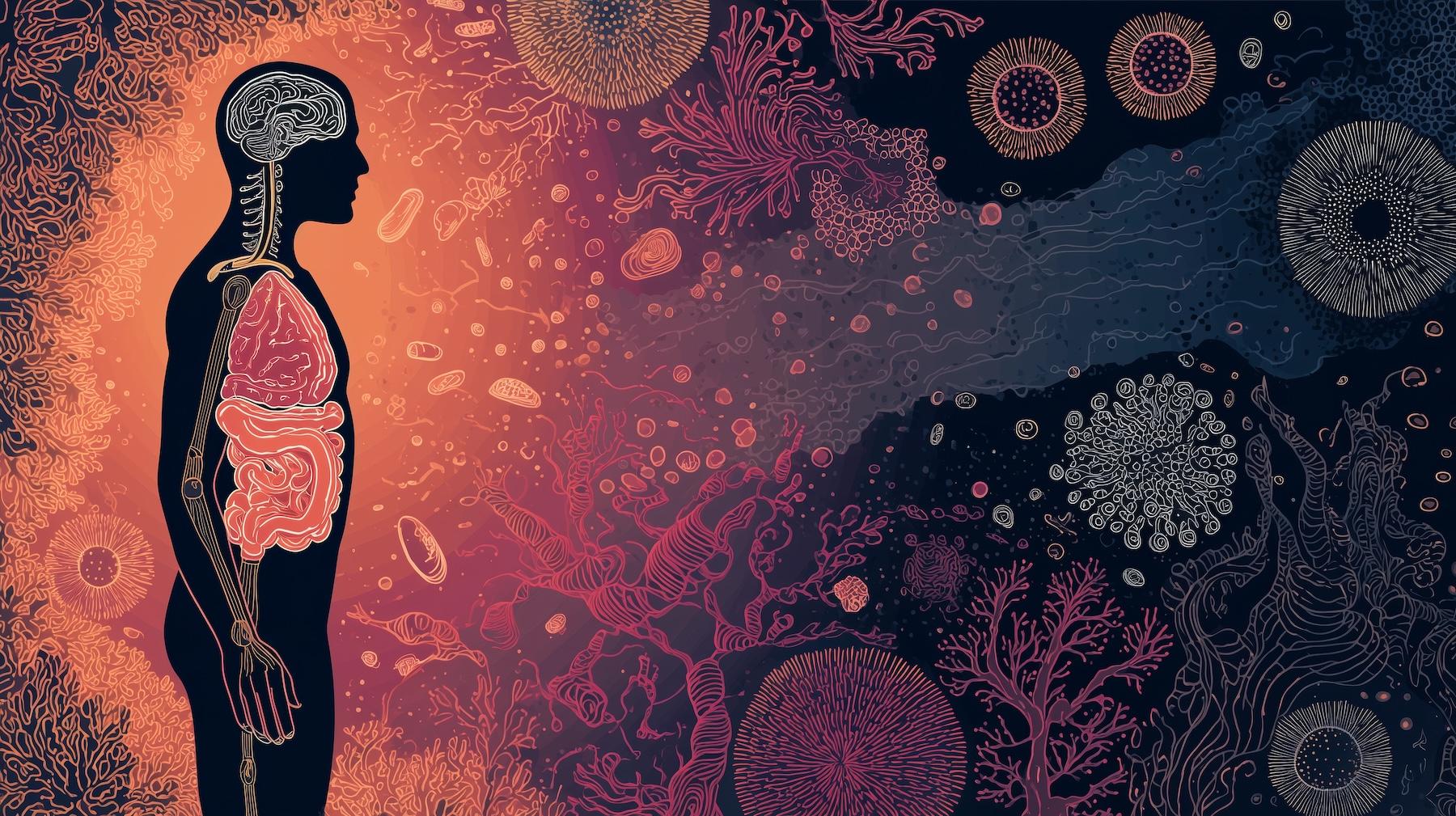
While the liver plays a significant role in systemic glutathione metabolism, the central nervous system (CNS) maintains its own tightly regulated GSH pool, independent of peripheral control (3-4). In the brain, glutathione helps maintain redox balance, reduce oxidative stress, and modulate immune signaling. It is most concentrated in cortical glial cells and microglia—key immune cells accounting for 10% of brain cells; both of which contribute to GSH synthesis and neuroimmune modulation (5-6).
Beyond direct antioxidant activity, glutathione functions through several enzymatic systems central to detoxification and redox control, including glutathione-S-transferases for conjugation reactions, glyoxylases, glutathione peroxidases and peroxiredoxins for peroxide reduction, and glutaredoxins for thiol-disulfide exchange. Through these integrated pathways, glutathione serves as a key regulator of brain cellular metabolism, supporting long-term neuronal function.
Key Mechanisms–Redox Balance, Detoxification, & Neuroprotection
Glutathione (GSH) governs cellular redox homeostasis and detoxifies reactive oxygen and nitrogen species (RONS), protecting neurons that are particularly vulnerable due to high metabolic demand and oxygen use. It also facilitates DNA synthesis and repair, stabilizes cellular membranes, and regulates appropriate immune responses (5-7).
As glutathione synthesis in the brain relies on cysteine availability, the neuronal transporter excitatory amino acid carrier 1 (EAAC1) plays a critical role in importing cysteine and glutamate. EAAC1 is tightly regulated by molecules, including glutamate transporter-associated protein 3–18 (GTRAP3-18) and miR-96-5p, offering potential therapeutic targets to optimize neuronal GSH levels and mitigate early neurodegeneration (6).
Glutathione synthesis is also supported by hepatic cysteine production via the transsulfuration pathway, which converts methionine to cysteine through homocysteine intermediates (7-8). GSH accounts for about 95% of the total non-protein thiols within cells, with intracellular concentrations ranging from 0.5 to 10 mM, far surpassing the levels of free cysteine (3-4).
Neuroprotection in the Central Nervous System
Glutathione (GSH) plays a vital neuroprotective role in the central nervous system (CNS), primarily by regulating redox homeostasis, detoxifying electrophilic compounds, and modulating excitotoxic signaling pathways. Its ability to scavenge reactive oxygen species (ROS) is especially critical given the CNS’s high metabolic activity and inherent susceptibility to oxidative stress-induced cellular injury.
Antioxidant defenses, including GSH synthesis and utilization, demonstrate regional heterogeneity across the brain. This variability is influenced by differences in the permeability of the blood–brain barrier (BBB) and the blood–cerebrospinal fluid (CSF) barrier, as well as by region-specific expression of enzymes involved in glutathione metabolism. For example, the cerebellum exhibits relatively high glutathione activity, which contributes to its enhanced resistance to oxidative damage. This neuroprotection is partly mediated by modulation of NMDA receptor activity and the maintenance of intracellular redox balance (9).
Ghersi-Egea et al. illustrated how the selective permeability of the blood-brain-barrier (BBB) and blood–CSF barrier, combined with the spatial distribution of glutathione-metabolizing enzymes, underlies region-specific vulnerability to oxidative injury in the CNS (9). Further supporting this, Lock and colleagues demonstrated that glutathione protects against 2-chloropropionic acid (CPA)-induced cerebellar toxicity by preserving thiol redox equilibrium and potentially interacting with NMDA receptor-mediated pathways.
In addition to its antioxidant function, GSH contributes to detoxification through its role in conjugation reactions catalyzed by glutathione S-transferases (GSTs), thereby preventing the accumulation of neurotoxic metabolites and supporting neuronal integrity. GSH also regulates redox-sensitive signaling pathways, modulates inflammatory processes, and supports innate immune responses—further underscoring its central role in CNS neuroprotection.
Overall, glutathione’s neuroprotective efficacy in the CNS is intricately regulated by enzyme expression, metabolic substrate availability, and regional brain biochemistry, making it a pivotal target for intervention in oxidative stress-related neuropathologies.
Glutathione & Stroke Resilience
Glutathione (GSH) plays a central role in protecting hippocampal neurons from ischemia-induced oxidative damage, a factor in post-stroke pathology. GSH acts by scavenging reactive oxygen species (ROS) and chelating intracellular zinc (Zn²⁺), whose dysregulation exacerbates neuronal injury and contributes to ischemic cascades (10). The rate-limiting step in GSH synthesis is cysteine availability, regulated predominantly by the excitatory amino acid carrier 1 (EAAC1) transporter in neurons. Experimental models demonstrate that upregulating EAAC1 function enhances cysteine uptake, elevates GSH levels, and significantly reduces neuronal apoptosis and infarct size following ischemic insult. Clinically, strategies aimed at modulating EAAC1-mediated cysteine transport and boosting endogenous GSH synthesis may hold promise for mitigating post-ischemic neurodegeneration and cognitive deficits associated with vascular dementia. Ongoing research into pharmacological agents or gene therapies targeting this pathway could translate into novel neuroprotective interventions in stroke management.
Neurodegenerative Disease & Aging
GSH levels decline with age and in neurodegenerative conditions, including Alzheimer’s, Parkinson’s, and Huntington’s disease, in addition to stroke. This depletion compromises the brain’s antioxidant defense, leading to increased oxidative damage and neuronal death. In Alzheimer’s disease models, oral glutathione supplementation improved cognition and reduced depressive-like behavior (11).
A recent 2022 study demonstrated that not only neurons but also glial cells rely on glutathione for homeostasis (12). A disrupted redox balance, particularly elevated oxidized glutathione (GSSG), is implicated in brain aging and neurodegeneration. Preserving its reduced state and supporting GSH levels may, therefore, promote healthy brain aging and enhance resilience to neurological disease.
A systematic review of 35 studies found consistent age-related GSH decline in blood and specific brain regions, including the precuneus, hippocampus, cingulate, and frontal cortex. While some MRS data showed region-specific increases, the overall trend pointed to rising oxidative stress with age (13). Notably, oxidized glutathione (GSSG) increased in areas, including the caudate nucleus, highlighting region-specific redox imbalances.
Immune Modulation & Neuroinflammation
In addition to its antioxidant effects, glutathione supports brain health by modulating systemic and neuroimmune responses. A pilot clinical study by Sasaninia et al. showed that topical glutathione–cyclodextrin (GSH-CD) increased intracellular GSH in mononuclear and red blood cells, decreased malondialdehyde (MDA), and significantly upregulated immune cytokines including IL-2, IFN-γ, IL-12p70, and TNF-α. (14). In vitro challenge with Mycobacterium avium demonstrated enhanced immune clearance post-treatment, suggesting that glutathione primes immune defense mechanisms relevant to neuroinflammatory conditions.
In a related study, Baker & Patel demonstrated that transdermal glutathione rapidly modulated systemic cytokines and decreased lipid peroxidation within hours (15). These immunomodulatory and antioxidant effects have direct implications for brain health, particularly in conditions involving neuroinflammation or oxidative stress. Importantly, endogenous GSH production begins to decline after age thirty, increasing vulnerability to environmental insults and chronic disease. A personalized approach for mitigating this decline with nutrition, lifestyle factors, and supplementation may help maintain redox balance.
Glutathione & Depression—Region-Specific Depletion in the Brain
Glutathione’s role in psychiatric disorders—particularly major depressive disorder (MDD)—has become increasingly evident as research into redox dysregulation and oxidative stress advances. A 2024 meta-analysis and systematic review by Bell and colleagues investigated brain glutathione levels in individuals with depression using proton magnetic resonance spectroscopy (¹H-MRS). Their findings contribute nuanced insight into the regional vulnerability of the brain’s antioxidant defense in the context of mood disorders (16).
Drawing on data from eight case-control studies comprising 230 individuals with depression and 216 healthy controls, the authors examined glutathione levels across two main brain regions: the occipital cortex and the medial frontal cortex. In the occipital cortex—a region associated with visual processing and also implicated in emotional regulation—glutathione concentrations were significantly lower in individuals with MDD compared to controls. The effect size was robust (Hedges’ g = –0.98), indicating a pronounced depletion in antioxidant capacity in this region.
In contrast, no statistically significant difference in glutathione levels was found in the medial frontal cortex, a brain region more directly tied to executive function, emotional processing, and self-referential thinking, highlighting the importance of analyzing glutathione’s role in depression on a region-specific basis.
These findings lend support to the hypothesis that high levels of oxidative stress may contribute to the pathophysiology of depression—though its effects may be anatomically selective. The results also underscore the limitations of current imaging methodologies and sample sizes, as the field still lacks high-quality, well-controlled magnetic resonance spectroscopy (MRS) studies that consistently assess glutathione in multiple brain areas. Nevertheless, the observed occipital depletion aligns with broader evidence of redox imbalance and impaired mitochondrial function in depressive disorders.
From a clinical perspective, these findings underscore the value of exploring interventions that modulate and support oxidative stress pathways—particularly those relative to glutathione synthesis or recycling—as adjunctive strategies for managing treatment-resistant depression. The region-specific pattern of glutathione loss may also help guide future neuroimaging studies and more effective targeted therapies.
Clinical Implications
Glutathione’s multifaceted roles in redox signaling, immune modulation, detoxification, and neurotransmitter regulation position it as a central mediator of brain health. Its functions in mitigating oxidative stress, supporting immune function, and modulating inflammation have widespread implications on health and the progression of disease.
Glutathione deficiency or dysregulation is a well-established biochemical hallmark in diverse chronic diseases, including neurodegenerative conditions, Parkinson’s, Alzheimer’s, mental health, cognitive decline, and metabolic and immune dysfunction, among others (17). These conditions share common mechanisms involving disrupted redox homeostasis, elevated oxidative stress, and impaired detoxification, underscoring the therapeutic potential of strategies aimed at restoring glutathione balance to mitigate disease progression.
Reliable imaging tools such as magnetic resonance spectroscopy (MRS) now allow real-time measurement of brain GSH levels, facilitating early detection and therapeutic monitoring. Whether via dietary precursors (e.g., N-acetylcysteine), targeted transdermal delivery, or enhancement of cysteine transport, interventions that optimize glutathione metabolism represent promising strategies for clinical brain health support.
Lifestyle modifications, including reducing toxin buildup (e.g., refraining from heavily processed foods, chemicals, and substances), while consuming whole nutrient-dense foods (e.g., phytonutrients, brassica veggies, and quality proteins, including rich sources of cysteine and selenium for GSH synthesis), getting adequate sleep, movement, sunshine and circadian rhythm balance, as well as mitigating stress are all valuable tools for supporting glutathione (17-21).
Join us for this compelling and relevant webinar, The Master Antioxidant: A Deep Dive into Glutathione’s Role in Metabolism, Mitochondrial Support, and Brain Function taking place on July 22nd from 5-7 PDT, featuring renowned experts: David Perlmutter, MD; Nayan Patel PharmD; and Jeffrey Bland, PhD as they further explore this robust molecule and its vast implications for our health.
References
- Vašková J, Kočan L, Vaško L, Perjési P. Glutathione-Related Enzymes and Proteins: A Review. 2023 Feb 2;28(3):1447. doi: 10.3390/molecules28031447. PMID: 36771108; PMCID: PMC9919958.
- Hopkins F.G. On glutathione: A reinvestigation. J. Biol. Chem. 1929;84:269–320. doi: 10.1016/S0021-9258(18)77062-2. [DOI] [Google Scholar]
- Pastore A., Federici G., Bertini E., Piemonte F. Analysis of glutathione: Implication in redox and detoxification. Chim. Acta. 2003;333:19–39. doi: 10.1016/S0009-8981(03)00200-6. [DOI] [PubMed] [Google Scholar]
- Wu G., Fang Y.Z., Yang S., Lupton J.R., Turner N.D. Glutathione metabolism and its implications for health. J. Nutr. 2004;134:489–492. doi: 10.1093/jn/134.3.489. [DOI] [PubMed] [Google Scholar]
- Commandeur J.N., Stijntjes G.J., Vermeulen N.P. Enzymes and transport systems involved in the formation and disposition of glutathione S-conjugates. Role in bioactivation and detoxication mechanisms of xenobiotics. Rev. 1995;47:271–330. [PubMed] [Google Scholar]
- Aoyama K. Glutathione in the Brain. Int J Mol Sci. 2021 May 9;22(9):5010. doi: 10.3390/ijms22095010. PMID: 34065042; PMCID: PMC8125908.
- Lu S.C. Regulation of hepatic glutathione synthesis: Current concepts and controversies. FASEB 1999;13:1169–1183. doi: 10.1096/fasebj.13.10.1169. [DOI] [PubMed] [Google Scholar]
- Mosharov E., Cranford M.R., Banerjee R. The quantitatively important relationship between homocysteine metabolism and glutathione synthesis by the transsulfuration pathway and its regulation by redox changes. 2000;39:13005–13011. doi: 10.1021/bi001088w. [DOI] [PubMed] [Google Scholar]
- Monks TJ, Ghersi-Egea JF, Philbert M, Cooper AJ, Lock EA. Symposium overview: the role of glutathione in neuroprotection and neurotoxicity. Toxicol Sci. 1999 Oct;51(2):161-77. doi: 10.1093/toxsci/51.2.161. PMID: 10543018.
- Higashi Y, Aratake T, Shimizu T, Shimizu S, Saito M. Protective Role of Glutathione in the Hippocampus after Brain Ischemia. Int J Mol Sci. 2021 Jul 21;22(15):7765. doi: 10.3390/ijms22157765. PMID: 34360532; PMCID: PMC8345998.
- Dwivedi D, Megha K, Mishra R, Mandal PK. Glutathione in Brain: Overview of Its Conformations, Functions, Biochemical Characteristics, Quantitation and Potential Therapeutic Role in Brain Disorders. Neurochem Res. 2020 Jul;45(7):1461-1480. doi: 10.1007/s11064-020-03030-1. Epub 2020 Apr 15. PMID: 32297027.
- Iskusnykh IY, Zakharova AA, Pathak D. Glutathione in Brain Disorders and Aging. 2022 Jan 5;27(1):324. doi: 10.3390/molecules27010324. PMID: 35011559; PMCID: PMC8746815.
- Detcheverry F, Senthil S, Narayanan S, Badhwar A. Changes in levels of the antioxidant glutathione in brain and blood across the age span of healthy adults: A systematic review. Neuroimage Clin. 2023;40:103503. doi: 10.1016/j.nicl.2023.103503. Epub 2023 Aug 26. PMID: 37742519; PMCID: PMC10520675.
- Sasaninia K, Kelley M, Abnousian A, Badaoui A, Alexander L, Sheren N, Owens J, Rajurkar S, Razo-Botello B, Chorbajian A, Yoon S, Dhama S, Avitia E, Ochoa C, Yutani R, Venketaraman V. Topical Absorption of Glutathione-Cyclodextrin Nanoparticle Complex in Healthy Human Subjects Improves Immune Response against Mycobacterium avium Antioxidants (Basel). 2023 Jul 2;12(7):1375. doi: 10.3390/antiox12071375. PMID: 37507915; PMCID: PMC10376088.
- Baker S. Nayan Patel, PharmD, Founder of Auro Wellness and CEO of Central Drugs Compounding Pharmacy. Integr Med (Encinitas). 2023 Sep;22(4):20-23. PMID: 37752928; PMCID: PMC10519235.
- Bell CJM, Mehta M, Mirza L, Young AH, Beck K. Glutathione alterations in depression: a meta-analysis and systematic review of proton magnetic resonance spectroscopy studies. Psychopharmacology (Berl). 2025 Apr;242(4):717-724. doi: 10.1007/s00213-024-06735-1. Epub 2024 Dec 21. PMID: 39708105; PMCID: PMC11890406.
- Minich DM, Brown BI. A Review of Dietary (Phyto)Nutrients for Glutathione Support. 2019 Sep 3;11(9):2073. doi: 10.3390/nu11092073. PMID: 31484368; PMCID: PMC6770193.
- Tkaczenko H, Kurhaluk N. Antioxidant-Rich Functional Foods and Exercise: Unlocking Metabolic Health Through Nrf2 and Related Pathways. Int J Mol Sci. 2025 Jan 27;26(3):1098. doi: 10.3390/ijms26031098. PMID: 39940866; PMCID: PMC11817741.
- Everson CA, Laatsch CD, Hogg N. Antioxidant defense responses to sleep loss and sleep recovery. Am J Physiol Regul Integr Comp Physiol. 2005 Feb;288(2):R374-83. doi: 10.1152/ajpregu.00565.2004. Epub 2004 Oct 7. PMID: 15472007.
- Chow ES, Long DM, Giebultowicz JM. Circadian rhythm in mRNA expression of the glutathione synthesis gene Gclc is controlled by peripheral glial clocks in Drosophila melanogaster. Physiol Entomol. 2016 Dec;41(4):369-377. doi: 10.1111/phen.12164. Epub 2016 Aug 26. PMID: 28503020; PMCID: PMC5423673.
- Maher P. The effects of stress and aging on glutathione metabolism. Ageing Res Rev. 2005 May;4(2):288-314. doi: 10.1016/j.arr.2005.02.005. PMID: 15936251.