Glutathione’s Role in Health: From Mitochondria to Metabolism
Glutathione (GSH) is one of the most critical molecular determinants of cellular resilience and systemic health. As a direct antioxidant, redox buffer, and essential enzymatic cofactor, GSH supports mitochondrial integrity, facilitates detoxification, modulates immune responses, and contributes to the repair of oxidative damage.
Although its physiological importance is well established, recent advances—described in the comprehensive 2024 review by Gasmi et al.—broaden our understanding of glutathione’s expanded roles in neuroimmune signaling, metabolic homeostasis, and chronic disease prevention (1). These emerging insights, discussed below, underscore the need to translate redox biochemistry into clinical application through targeted interventions that restore and sustain glutathione function in the context of aging, neurodegeneration, immune dysfunction, and environmental toxin burden.
A Deeper Dive Into This Master Antioxidant
GSH is a vital thiol-containing tripeptide present in nearly all living cells, where it serves multiple overlapping roles in maintaining cellular integrity and physiological homeostasis. In addition to its central function as an intracellular antioxidant, it serves as a significant cofactor for several antioxidant enzymes—including glutathione peroxidase (GPx) and glutathione S-transferases (GSTs)—that neutralize reactive oxygen species (ROS) and other electrophilic compounds (1–4). GSH also helps maintain redox homeostasis by supporting other antioxidant systems, further underscoring its central role in cellular defense.
Structure, Biosynthesis, and Reversible Redox Cycling
GSH is a tripeptide (L-γ-glutamyl-L-cysteinyl-glycine) synthesized intracellularly in a two-step ATP-dependent process. Glutamate and cysteine form γ-glutamylcysteine, followed by the addition or conjugation of glycine to complete GSH synthesis (1-3). The cysteine residue contains a reactive thiol (-SH) group integral for glutathione’s antioxidant, redox, and conjugation functions (4-5).
Glutathione exists in two primary forms: the reduced form (GSH), characterized by a free sulfhydryl group (-SH) on its cysteine residue, and the oxidized form, glutathione disulfide (GSSG), which is formed when two GSH molecules are linked via a disulfide bond between their cysteine residues during redox reactions. This reversible redox cycling between GSH and GSSG is integral for neutralizing free radicals and maintaining cellular redox homeostasis (6).
Cellular Metabolism & Localization
Beyond its antioxidant functions, GSH contributes directly to cellular metabolism. Certain metabolic intermediates derived from GSH catabolism can enter the tricarboxylic acid (TCA) cycle, thereby supporting mitochondrial ATP production (7). Furthermore, GSH plays an essential role in the glutathione-ascorbate cycle, facilitating the reduction of hydrogen peroxide (H₂O₂)—a highly reactive oxidant that, if not neutralized, can damage lipids, proteins, and DNA (4–8).
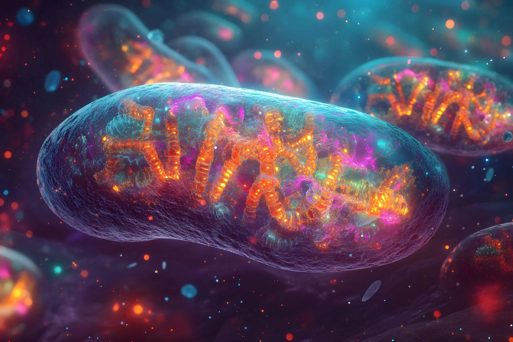
GSH is found predominantly in the cytosol (~90%), with smaller but vital pools in mitochondria and the endoplasmic reticulum. Mitochondrial GSH is chiefly important for detoxifying ROS generated through oxidative phosphorylation (6-8). Depletion of mitochondrial GSH is associated with cellular senescence, compromised energy production, and age-related pathology (9–10).
Intracellular concentrations range from 1–2 mM in most cells to ~10 mM in hepatocytes, which also export GSH into plasma and bile. Although plasma levels are typically in the micromolar range, epithelial surfaces (e.g., lungs) maintain high extracellular GSH concentrations to protect against inhaled oxidants, such as hypochlorous acid (HOCl) (11-14).
BIOCHEMICAL FUNCTIONS & CLINICAL RELEVANCE OF GLUTATHIONE
Antioxidant Defense & Redox Regulation
GSH is the most abundant intracellular antioxidant, neutralizing reactive oxygen (ROS), reactive nitrogen (RNS), and reactive sulfur (RSS) species (5-6). Through redox cycling, GSH donates electrons to quench free radicals and is then oxidized to glutathione disulfide (GSSG). Glutathione reductase regenerates GSH utilizing NADPH, maintaining a dynamic antioxidant buffer (6).
This mechanism protects thiol groups in proteins, stabilizes membrane lipids, and preserves DNA integrity. In healthy cells, more than 98% of GSH remains in its reduced form, with a typical GSH:GSSG ratio of above 100:1. Oxidative stress can reduce this ratio to 10:1 or lower, making the redox couple a sensitive marker of cellular oxidative status (6, 15).
Regeneration of Non-Enzymatic Antioxidants
GSH not only neutralizes reactive species directly, but also helps regenerate oxidized antioxidants, including vitamin C and polyphenols (quercetin, resveratrol), enhancing overall ROS-scavenging capacity (9, 16-17).
Polyphenols also stimulate phase II detoxification enzymes (glutathione-S-transferases) and cytochrome P450 systems, which depend on GSH for proper function (18-19).
Water-soluble antioxidants (GSH, vitamin C) are active in the cytosol and plasma, while lipid-soluble antioxidants (vitamin E, carotenoids) localize to membranes (20). Sulfur-rich plasma proteins, including albumin and ceruloplasmin, further buffer redox stress by chelating metal ions and exhibiting thiol-based antioxidant effects (21).
Alpha-lipoic acid (α-LA) enhances GSH synthesis by activating the Nrf2 signaling pathway, a transcriptional regulator of antioxidant genes. Inhibition of Nrf2 significantly reduces GSH biosynthetic enzyme expression (22). α-LA has shown promise in diabetes, cardiovascular disease, and metabolic syndrome due to its mitochondrial, anti-inflammatory, and insulin-mimetic effects (23-24).
Role of Glutathione in Detoxification
GSH is fundamental in neutralizing harmful endogenous and exogenous substances, including xenobiotics, reactive metabolites, and environmental toxins. It functions as a key cellular antioxidant, undergoing oxidation by glutathione peroxidase (GPx) and regeneration via glutathione reductase using NADPH (25).
Hepatic Detoxification via Glutathione-S-Transferases – In hepatocytes—the primary site of xenobiotic metabolism—glutathione (GSH) conjugates electrophilic toxins via glutathione-S-transferases (GSTs), enhancing their solubility to facilitate elimination through bile or urine (26). GST isoforms (GSTM1, GSTP1, GSTT1) are expressed in the cytosol, mitochondria, and nucleus across multiple organs, including the liver, kidneys, lungs, brain, and placenta, exhibiting broad substrate specificity against electrophiles (molecules deficient in electron pairs) and ROS (27–33).
Phase I-III Metabolic Detoxification Cascade – Detoxification proceeds via three enzymatic phases: Phase I enzymes (cytochrome P450 oxidases) introduce reactive groups to xenobiotics; Phase II enzymes, such as GSTs, conjugate these metabolites with endogenous molecules like GSH, neutralizing toxicity; and Phase III transporters export conjugates for excretion.
Regulation & Induction of Detoxifying Enzymes – GST expression is upregulated in response to oxidative and proliferative stressors, including those associated with tumorigenesis. These enzymes play a critical role in cellular defense by catalyzing the conjugation of electrophilic compounds with glutathione, thereby facilitating the neutralization of carcinogens, reduction of lipid peroxides, regeneration of oxidized thiol groups, and repair of damaged membrane phospholipids (28–30). Natural bioactive compounds (polyphenols) enhance detoxification capacity by upregulating GST expression and promoting glutathione biosynthesis (34–36).
Mitochondrial Protection & Sex Differences in GSH Metabolism – GSH synergizes with ascorbate (vitamin C) to protect mitochondria from oxidative injury. Studies show that female rodents have higher mitochondrial GSH levels and antioxidant enzyme activities, linked to estrogen-mediated activation of MAPK and NF-κB signaling pathways, suggesting sex-based differences in redox resilience (37-39).
Xenobiotic-Induced Oxidative Stress & Cellular Signaling – Reactive oxygen and nitrogen species generated by xenobiotic metabolism can cause DNA damage, protein oxidation, and lipid peroxidation if not adequately regulated; however also serve as critical intracellular messengers modulating cell survival adaptive responses and pathways (40).
Nrf2/Keap1/ARE Redox Regulatory Pathway – In response to oxidative or xenobiotic stress, Nrf2 dissociates from its inhibitor Keap1, translocates to the nucleus, and binds antioxidant response elements (ARE) to induce expression of genes involved in antioxidant defense and GSH biosynthesis (41).
Systemic Impact of GSH Detoxification – The GSH system detoxifies over 300 oxidized or electrophilic compounds, including reactive oxygen intermediates, aldehydes, and environmental toxins, converting them into excretable, biologically inert forms (42). It also neutralizes toxins in the GI tract prior to systemic absorption and clears absorbed toxins via hepatic and renal pathways. Additionally, GSH enhances immune defense by activating natural killer (NK) cells and promoting T-lymphocyte proliferation (42).
Roles in Cell Cycle, Senescence, & Apoptosis
GSH modulates key cellular processes, including proliferation, senescence, and apoptosis (1). Nuclear GSH concentrations (~0.7 mM) modulate telomerase activity and protect DNA from oxidative damage (43). Disrupted GSH homeostasis has been linked to pathologies including cancer, neurodegenerative disorders, and immune dysfunction (44–46).
Neuroprotection – GSH is vital for redox balance in the central nervous system. Astrocytes supply GSH precursors to neurons, enabling detoxification of ROS generated during high metabolic activity (44, 47). Adequate GSH levels help preserve neuronal integrity, while depletion is a hallmark of neurodegenerative diseases such as Parkinson’s and Alzheimer’s (48–49).
Cancer Biology & Drug Resistance – GSH plays a complex, context-dependent role in cancer biology. Under physiological conditions, it contributes to genomic stability by detoxifying carcinogens and reactive metabolites. However, elevated GSH levels have been reported in multiple malignancies—including those of the bone marrow, breast, colon, larynx, and lung—and are associated with tumor progression and increased metastatic potential (50).
In cancer cells, high intracellular GSH enhances resistance to oxidative stress and reduces the cytotoxicity of chemotherapeutic agents. This effect is mediated by GSH-dependent conjugation and export of anticancer drugs, diminishing their intracellular concentrations and therapeutic efficacy (50–53). While GSH is essential for normal redox homeostasis, its upregulation in tumor cells contributes to chemoresistance and poses a challenge to effective cancer treatment.
Routes of Administration
GSH can be administered via oral, sublingual, intravenous (IV), intramuscular (IM), inhalational, and transdermal routes. Each delivery method differs in bioavailability, systemic absorption, and tissue distribution, with route selection guided by the therapeutic goal and clinical context (1).
Inhaled GSH—delivered via aerosolized or nebulized formulations—has been studied for respiratory conditions such as cystic fibrosis and idiopathic pulmonary fibrosis, owing to its localized antioxidant activity within the pulmonary epithelium (54). Intramuscular (IM) GSH has been utilized in clinical settings to support systemic redox balance in patients undergoing chemotherapy, individuals with HIV/AIDS, and in the treatment of male infertility (55). Intravenous GSH offers rapid systemic bioavailability and has demonstrated hepatoprotective properties in preclinical studies, including protection against ischemia-reperfusion injury during liver transplantation (56). Its favorable safety profile further supports its potential as an adjunctive therapy in oxidative stress-related disorders.
Clinical & Nutritional Relevance
Given its diverse physiological functions, GSH is increasingly recognized for its potential in supporting health, aging, cancer, respiratory conditions, and neurodegenerative diseases with thoughtful utilization (44, 46).
Strategies to restore and enhance GSH levels include precursor supplementation—such as N-acetylcysteine (NAC), glycine, and methylated B vitamins—and lifestyle interventions that promote endogenous synthesis and recycling. These include reducing stress and toxicant exposure, ensuring adequate sleep, engaging in regular physical activity, and consuming a nutrient-dense diet rich in sulfur-containing vegetables, glycine-rich proteins, and vitamin C to support systemic redox balance.
GSH represents a fundamental pillar of cellular health with profound clinical relevance, impacting nearly every physiological system of the body (1).
Join us for this webinar, The Master Antioxidant: A Deep Dive into Glutathione’s Role in Metabolism, Mitochondrial Support, and Brain Function on July 22nd, from 5-7 PM PDT where leading experts Jeffrey Bland, PhD, David Perlmutter, MD, and Nayan Patel, PharmD further explore the clinical functions of glutathione in regulating metabolic efficiency, mitochondrial health, and neurophysiological function.
References
- Gasmi A, Nasreen A, Lenchyk L, Lysiuk R, Peana M, Shapovalova N, Piscopo S, Komisarenko M, Shanaida M, Smetanina K, Antonyak H, Fira L, Lykhatskyi P, Fira D, Bjørklund G. An Update on Glutathione’s Biosynthesis, Metabolism, Functions, and Medicinal Purposes. Curr Med Chem. 2024;31(29):4579-4601. doi: 10.2174/0109298673251025230919105818. PMID: 37921175.
- Lushchak, V.I. Glutathione homeostasis and functions: Potential targets for medical interventions. Amino Acids, 2012, 2012, 1-26. http://dx.doi.org/10.1155/2012/736837 PMID: 22500213
- Meister, A.; Anderson, M.E. Glutathione. Rev. Biochem., 1984, 52, 711-760.
- Forman, H.J.; Zhang, H.; Rinna, A. Glutathione: Overview of its protective roles, measurement, and biosynthesis. Aspects Med., 2009, 30(1-2), 1-12. http://dx.doi.org/10.1016/j.mam.2008.08.006 PMID: 1879631
- Vašková, J.; Kočan, L.; Vaško, L.; Perjési, P. Glutathionerelated enzymes and proteins: A review. Molecules, 2023, 28(3), 1447. http://dx.doi.org/10.3390/molecules28031447 PMID: 36771108
- Pizzorno, J. Glutathione! Med., 2014, 13(1), 8-12. PMID: 26770075
- Korost, Y.V.; Sokurenko, O.O.; Kalashchenko, S.I.; Odynetsʹ, M.O. Understanding biochemical processes as the proposal of successful disease treatment. State-Art Technol. Med., 2016, 3-4(129-130), 20-23.
- Marí, M.; de Gregorio, E.; de Dios, C.; Roca-Agujetas, V.; Cucarull, B.; Tutusaus, A.; Morales, A.; Colell, A. Mitochondrial glutathione: Recent insights and role in disease. Antioxidants, 2020, 9(10), 909.
- Forman, H.J.; Thomas, M.J. Oxidant production and bactericidal activity of phagocytes. Rev. Physiol., 1986, 48(1), 669-680. http://dx.doi.org/10.1146/annurev.ph.48.030186.003321 PMID: 3010830 ;
- Gao, X.; Yu, X.; Zhang, C.; Wang, Y.; Sun, Y.; Sun, H.; Zhang, H.; Shi, Y.; He, X. Telomeres and mitochondrial metabolism: Implications for cellular senescence and agerelated diseases. Stem Cell Rev. Rep., 2022, 18(7), 23152327. http://dx.doi.org/10.1007/s12015-022-10370-8 PMID: 35460064
- Roger, L.; Tomas, F.; Gire, V. Mechanisms and regulation of cellular senescence. Int. J. Sci., 2021, 22(23), 13173. http://dx.doi.org/10.3390/ijms222313173 PMID: 34884978
- Martini, H.; Passos, J. Cellular senescence: All roads lead to mitochondria. FEBS J., 2022, 290(5), 1186-1202. PMID: 35048548
- Meister, A. Glutathione metabolism and its selective modification. Biol. Chem., 1988, 263(33), 17205-17208. http://dx.doi.org/10.1016/S0021-9258(19)77815-6 PMID: 3053703
- Cantin, A.M.; North, S.L.; Hubbard, R.C.; Crystal, R.G. Normal alveolar epithelial lining fluid contains high levels of glutathione. J Appl Physiol, 1987, 63(1), 152-157.
- Ruiz-Capillas, C., Eds.; Nollet., L.M.L., Eds.; Flow Injection Analysis of Food Additives; Shpigun, L.K.CRC Press: C.Ruiz-Capillas, 2015, p. 736.
- Gasmi, A.; Mujawdiya, P.K.; Noor, S.; Lysiuk, R.; Darmohray, R.; Piscopo, S.; Lenchyk, L.; Antonyak, H.; Dehtiarova, K.; Shanaida, M.; Polishchuk, A.; Shanaida, V.; Peana, M.; Bjørklund, G. Polyphenols in metabolic diseases. Molecules., 2022, 27(19), 6280. http://dx.doi.org/10.3390/molecules27196280 PMID: 36234817
- Quispe, C.; Cruz-Martins, N.; Manca, M.L.; Manconi, M.; Sytar, O.; Hudz, N.; Shanaida, M.; Kumar, M.; Taheri, Y.; Martorell, M.; Sharifi-Rad, J.; Pintus, G.; Cho, W.C. Nanoderived therapeutic formulations with curcumin in inflammation-related diseases. Med. Cell. Longev., 2021, 2021, 1-15.
- Bjørklund, G.; Dadar, M.; Chirumbolo, S.; Lysiuk, R. Flavonoids as detoxifying and pro-survival agents: What’s new? Food Chem. Toxicol., 2017, 110, 240-250. http://dx.doi.org/10.1016/j.fct.2017.10.039 PMID: 29079495
- Chirumbolo, S.; Bjørklund, G.; Lysiuk, R.; Vella, A.; Lenchyk, L.; Upyr, T. Targeting cancer with phytochemicals via their fine tuning of the cell survival signaling pathways. J. Mol. Sci., 2018, 19(11), 3568. http://dx.doi.org/10.3390/ijms19113568 PMID: 30424557
- Valko, M.; Rhodes, C.J.; Moncol, J.; Izakovic, M.; Mazur, M. Free radicals, metals and antioxidants in oxidative stress-induced cancer. Biol. Interact., 2006, 160(1), 1-40. http://dx.doi.org/10.1016/j.cbi.2005.12.009 PMID: 16430879
- Babula, P.; Masarik, M.; Adam, V.; Eckschlager, T.; Stiborova, M.; Trnkova, L.; Skutkova, H.; Provaznik, I.; Hubalek, J.; Kizek, R. Mammalian metallothioneins: Properties and functions. Metallomics, 2012, 4(8), 739-750. http://dx.doi.org/10.1039/c2mt20081c PMID: 22791193
- Zhang, J.; Zhou, X.; Wu, W.; Wang, J.; Xie, H.; Wu, Z. Regeneration of glutathione by α-lipoic acid via Nrf2/ARE signaling pathway alleviates cadmium-induced HepG2 cell toxicity. Environ. Pharmacol., 2017, 51, 30-37. http://dx.doi.org/10.1016/j.etap.2017.02.022 PMID: 28262510
- Patel, J.; Matnor, N.A.; Iyer, A.; Brown, L. A regenerative antioxidant protocol of vitamin E and α-lipoic acid ameliorates cardiovascular and metabolic changes in fructose-fed rats. Evid. Based Complement. Med., 2011, 2011, 1-8. http://dx.doi.org/10.1155/2011/120801 PMID: 21437191
- Rochette, L.; Ghibu, S.; Muresan, A.; Vergely, C. Alphalipoic acid: Molecular mechanisms and therapeutic potential in diabetes. J. Physiol. Pharmacol., 2015, 93(12), 1021-1027. http://dx.doi.org/10.1139/cjpp-2014-0353 PMID: 26406389
- Potęga, A. Glutathione-mediated conjugation of anticancer drugs: An overview of reaction mechanisms and biological significance for drug detoxification and bioactivation. Molecules., 2022, 27(16), 5252. http://dx.doi.org/10.3390/molecules27165252 PMID: 36014491
- Tymoshenko, M.; Gaida, I.; Kravchenko, O.; Ostapchenko, I. Content of different forms of glutathione and activity of glutathione reductase in cells of the gastrointestinal mucosa under experimental gastric carcinogenesis [in Ukrainian]. Visnyk Kyyivsʹkoho Natsionalʹnoho Universytetu Imeni T. Shevchenka, 2012, 61, 34-36.
- Hayes, J.D. Glutathione transferases. Annu. Rev. Pharmacol. Toxicol., 2005, 45, 51-88.
- Chorna, I.V.; Dronik, G.V.; Rogozinsky, M.S. Biological values of antioxidant protection system indicators in assessing the safety of the exercise of genetically modified organisms. Young Scientist, 2018, 10(62), 461-469.
- Hayes, J.D.; Pulford, D.J. The glutathione S-transferase supergene family: Regulation of GST and the contribution of the isoenzymes to cancer chemoprotection and drug resistance. Rev. Biochem. Mol. Biol., 1995, 30(6), 445520. http://dx.doi.org/10.3109/10409239509083491 PMID: 8770536
- Cahill, L.E.; Fontaine-Bisson, B.; El-Sohemy, A. Functional genetic variants of glutathione S-transferase protect against serum ascorbic acid deficiency. J. Clin. Nutr., 2009, 90(5), 1411-1417. http://dx.doi.org/10.3945/ajcn.2009.28327 PMID: 19710200
- Xu, S.; Wang, Y.; Roe, B.; Pearson, W.R. Characterization of the human class Mu glutathione S-transferase gene cluster and the GSTM1 deletion. Biol. Chem., 1998, 273(6), 3517-3527. http://dx.doi.org/10.1074/jbc.273.6.3517 PMID: 9452477
- Frova, C. Glutathione transferases in the genomics era: New insights and perspectives. Eng., 2006, 23(4), 149169. http://dx.doi.org/10.1016/j.bioeng.2006.05.020 PMID: 16839810
- Makarchuk, V.A.; Ushakova, G.O.; Krylova, O.O. The glutathione system in the blood of rats and morphological changes of the pancreas under experimental acute and chronic pancreatitis. Biochem. J., 2013, 85(1), 71-78.
- Gons’kyĭ, IaI.; Korda, M.M.; Klishch, I.M. Status of the free radical oxidation and antioxidant system in rats with toxic liver damage; effect of tocopherol and dimethylsulfoxide. Biokhim. Zh., 1991, 63(5), 112-116. PMID: 1788866
- Skakun, N.P.; Stepanova, Y.N. Comparative evaluation of the hepatoprotective, antioxidant and choleretic activity of flavonoid drugs. Delo, 1988, 12, 52-54. PMID: 3245169
- Bjørklund, G.; Shanaida, M.; Lysiuk, R.; Butnariu, M.; Peana, M.; Sarac, I.; Strus, O.; Smetanina, K.; Chirumbolo, S. Natural compounds and products from an anti-aging perspective. Molecules, 2022, 27(20), 7084. http://dx.doi.org/10.3390/molecules27207084 PMID: 36296673
- Borras, C.; Gambini, J.; Vina, J. Mitochondrial oxidant generation is involved in determining why females live longer than males. Front Biosci., 2007, 12, 1008-1013.
- Kulinsky, V.I.; Kolesnichenko, L.S. Mitochondrial glutathione. Biochemistry., 2007, 72(7), 698-701. http://dx.doi.org/10.1134/S0006297907070024 PMID: 17680760
- Wang, L.; Ahn, Y.J.; Asmis, R. Sexual dimorphism in glutathione metabolism and glutathione-dependent responses. Redox Biol., 2020, 31, 101410. http://dx.doi.org/10.1016/j.redox.2019.101410 PMID: 31883838
- Lushchak, V.I. Free radicals, reactive oxygen species, oxidative stress and its classification. Biol. Interact., 2014, 224, 164-175. http://dx.doi.org/10.1016/j.cbi.2014.10.016 PMID: 25452175
- Bjørklund, G.; Dadar, M.; Martins, N.; Chirumbolo, S.; Goh, B.H.; Smetanina, K.; Lysiuk, R. Brief challenges on medicinal plants: An eye-opening look at ageing-related disorders. Basic Clin. Pharmacol. Toxicol., 2018, 122(6), 539-558. http://dx.doi.org/10.1111/bcpt.12972 PMID: 29369521
- Tkach, S.M. Glutathione as a universal hepatoprotector with pleiotropic effects. Health Ukraine, 2018, 2(48), 16-17.
- Kulinsky, V.I.; Kolesnichenko, L.S. Nuclear glutathione and its functions. Khim., 2010, 56(6), 657-662. http://dx.doi.org/10.18097/PBMC20105606657 PMID: 21395068
- Iskusnykh, I.Y.; Zakharova, A.A.; Pathak, D. Glutathione in brain disorders and aging. , 2022, 27(1), 324. http://dx.doi.org/10.3390/molecules27010324 PMID: 35011559
- Hajam, Y.A.; Rani, R.; Ganie, S.Y.; Sheikh, T.A.; Javaid, D.; Qadri, S.S.; Pramodh, S.; Alsulimani, A.; Alkhanani, M.F.; Harakeh, S.; Hussain, A.; Haque, S.; Reshi, M.S. Oxidative stress in human pathology and aging: Molecular mechanisms and perspectives. , 2022, 11(3), 552. http://dx.doi.org/10.3390/cells11030552 PMID: 35159361
- Tew, K.D. Glutathione-associated enzymes in anticancer drug resistance. Cancer Res., 2016, 76(1), 7-9. http://dx.doi.org/10.1158/0008-5472.CAN-15-3143 PMID: 26729789
- Ennis, S.R.; Kawai, N.; Ren, X.; Abdelkarim, G.E.; Keep, R.F. Glutamine uptake at the blood-brain barrier is mediated by N-system transport. Neurochem., 1998, 71(6), 2565-2573. http://dx.doi.org/10.1046/j.1471-4159.1998.71062565.x PMID: 9832157
- Aoyama, K. Glutathione in the Brain. Int. J. Mol. Sci., 2021, 22(9), 5010.
- Bjørklund, G.; Peana, M.; Maes, M.; Dadar, M.; Severin, B. The glutathione system in Parkinson’s disease and its progression. Biobehav. Rev., 2021, 120, 470-478. http://dx.doi.org/10.1016/j.neubiorev.2020.10.004 PMID: 33068556
- Bansal, A.; Simon, M.C. Glutathione metabolism in cancer progression and treatment resistance. Cell Biol., 2018, 217(7), 2291-2298. http://dx.doi.org/10.1083/jcb.201804161 PMID: 29915025
- Kurutas, E.B. The importance of antioxidants which play the role in cellular response against oxidative/nitrosative stress: current state. J., 2015, 15(1), 71. http://dx.doi.org/10.1186/s12937-016-0186-5 PMID: 27456681
- Lobo, V.; Patil, A.; Phatak, A.; Chandra, N. Free radicals, antioxidants and functional foods: Impact on human health. Rev., 2010, 4(8), 118-126. http://dx.doi.org/10.4103/0973-7847.70902 PMID: 22228951
- Estrela, J.M.; Ortega, A.; Obrador, E. Glutathione in cancer biology and therapy. Crit. Clin. Lab. Sci., 2006, 43(2), 143-181. http://dx.doi.org/10.1080/10408360500523878 PMID: 16517421
- Prousky, J. The treatment of pulmonary diseases and respiratory-related conditions with inhaled (nebulized or aerosolized) glutathione. Based Complement. Alternat. Med., 2008, 5(1), 27-35. http://dx.doi.org/10.1093/ecam/nem040 PMID: 18317545
- Palamara, A.T.; Garaci, E.; Rotilio, G.; Ciriolo, M.R.; Casablanca, A.; Fraternale, A.; Rossi, L.; Schiavano, G.F.; Chiarantlni, L.; Magnani, M. Inhibition of murine AIDS by reduced glutathione. AIDS Res. Hum. Retroviruses, 1996, 12(14), 1373-1381.
- Schauer, R.J.; Kalmuk, S.; Gerbes, A.L.; Leiderer, R.; Meissner, H.; Schildberg, F.W.; Messmer, K.; Bilzer, M. Intravenous administration of glutathione protects parenchymal and non-parenchymal liver cells against reperfusion injury following rat liver transplantation. World J. Gastroenterol., 2004, 10(6), 864-870. http://dx.doi.org/10.3748/wjg.v10.i6.864 PMID: 15040034