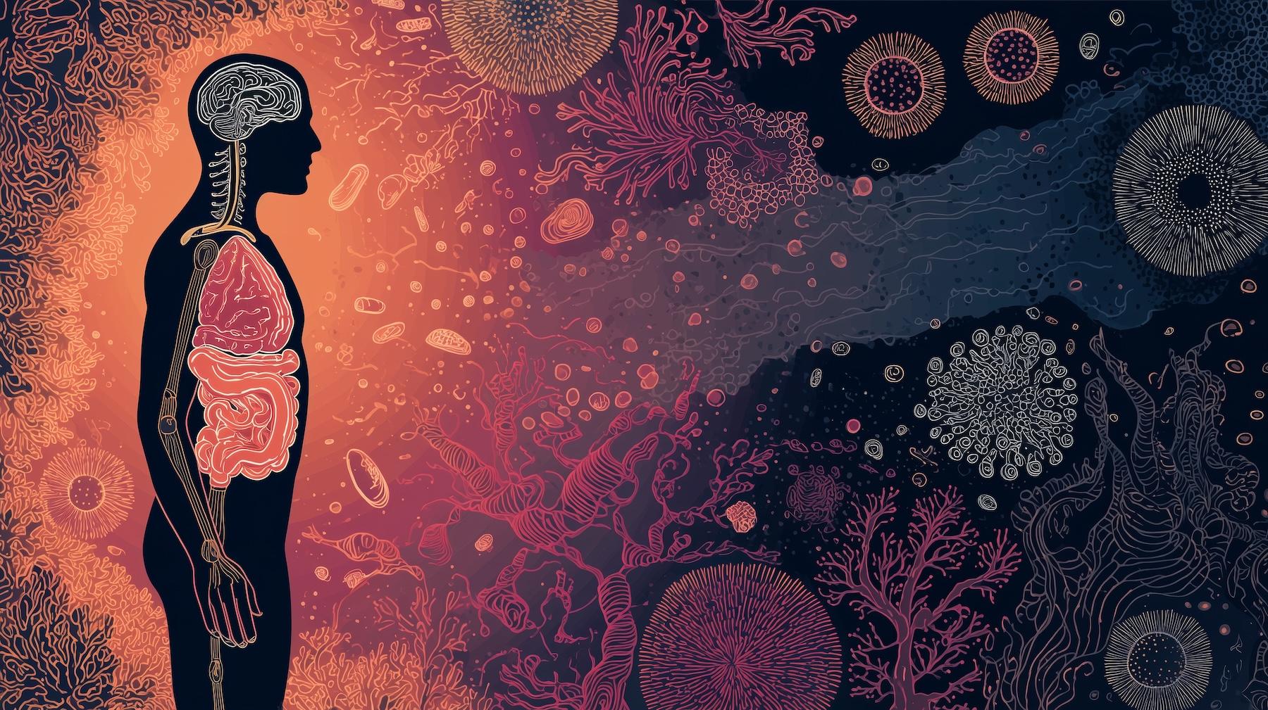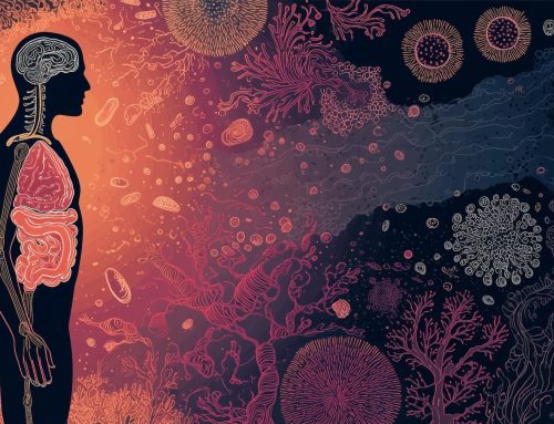
The Systems Biology of Glutathione and Mitochondrial Stress in Cardiometabolic Risk
Glutathione sits at the core of cellular redox homeostasis. As the most abundant intracellular antioxidant, reduced glutathione (GSH) functions not merely as a scavenger of reactive oxygen species (ROS), but as a pivotal modulator of redox-sensitive enzymatic activity, mitochondrial integrity, inflammatory signaling, and metabolic resilience and flexibility. When glutathione synthesis or recycling becomes impeded, the resulting redox imbalance initiates a cascade of oxidative damage that disproportionately affects the vascular endothelium and cardiometabolic systems.
Mitochondrial stress accelerates and amplifies these pathological processes, creating a self-reinforcing cycle of oxidative injury, impaired bioenergetics, and cardiometabolic dysfunction (1-2). These interconnected processes reveal how even subtle impairments in glutathione synthesis can cascade into widespread oxidative damage, setting the stage for vascular and metabolic dysfunction.
Impaired Glutathione Synthesis & Recycling in Cellular Redox Homeostasis
To understand these systemic consequences, it is essential to examine the tightly regulated biochemical pathways that govern glutathione synthesis and recycling.
Glutathione is a tripeptide composed of glutamate, cysteine, and glycine, synthesized intracellularly through a tightly regulated, ATP-dependent pathway. Of these substrates, cysteine availability is rate-limiting, rendering glutathione synthesis particularly sensitive to dietary protein quality, sulfur amino acid intake, and intracellular redox demand. Once oxidized during ROS detoxification (most notably through glutathione peroxidase–mediated reduction of hydrogen peroxide), glutathione must be regenerated from its oxidized form (GSSG) by glutathione reductase, a process dependent on a continuous supply of NADPH from the pentose phosphate pathway (3).
Under physiological conditions, cells maintain a highly reduced intracellular environment, reflected in a GSH: GSSG ratio near 100:1 (4-5). Disruption at any point—substrate limitation, enzymatic insufficiency, impaired NADPH availability, or excessive oxidative burden—drives this ratio downward, shifting cells toward an oxidized redox state. Loss of redox flexibility compromises protein thiol regulation, redox-sensitive signaling pathways, and endothelial homeostasis, positioning glutathione insufficiency as a primary upstream disturbance rather than a downstream consequence of disease (1-3).
Mitochondrial compartments are particularly sensitive to these shifts. Mitochondrial glutathione pools are indispensable for maintaining respiratory chain function, preventing lipid peroxidation, and regulating apoptotic signaling (1). When mitochondrial glutathione becomes depleted, oxidative damage accelerates at the primary site of ROS generation.
Oxidative Stress as the Link to Vascular Dysfunction
Disruption of glutathione homeostasis has immediate implications for the vascular endothelium, where oxidative stress directly undermines nitric oxide (NO) signaling and vascular integrity.
The vascular endothelium is uniquely vulnerable to glutathione insufficiency. Endothelial cells are continuously exposed to shear stress, circulating glucose and lipids, and inflammatory mediators, all of which generate ROS. Adequate intracellular glutathione is therefore essential for neutralizing oxidants and preserving nitric oxide (NO) bioavailability.
When glutathione synthesis or recycling is compromised, hydrogen peroxide and lipid peroxides accumulate, oxidizing endothelial membranes and proteins (6). This oxidative injury disrupts endothelial nitric oxide synthase (eNOS) function, reduces nitric oxide (NO) bioavailability, and promotes vascular stiffness—an early mechanistic link between redox imbalance and endothelial dysfunction. In parallel, redox-sensitive transcription factors such as NF-κB are activated, upregulating adhesion molecules, chemokines, and pro-inflammatory cytokines (7).
Over time, repeated oxidative injury promotes endothelial apoptosis and maladaptive repair responses, including smooth muscle proliferation and vascular calcification. Mitochondrial glutathione depletion has emerged as a key determinant of endothelial vulnerability, directly linking mitochondrial redox failure to vascular pathology (2,8).
Cardiometabolic Imbalance & Glutathione Insufficiency
Endothelial oxidative injury intersects with metabolic tissues, compounding insulin resistance, dyslipidemia, and systemic inflammation.
The cardiometabolic consequences of impaired glutathione homeostasis are profound. In insulin-sensitive tissues, oxidative stress interferes with insulin receptor signaling through modification of critical cysteine residues and activation of stress kinases. In type 2 diabetes, glutathione depletion exacerbates hyperglycemia-induced ROS production, creating a feed-forward loop in which oxidative stress both results from and worsens insulin resistance (9).
Elevated glucose flux increases mitochondrial electron pressure, driving excess superoxide generation. In the absence of sufficient glutathione buffering, oxidative damage accumulates in pancreatic β-cells, hepatocytes, adipocytes, and vascular tissues. These changes impede glucose disposal, promote dyslipidemia, and sustain chronic low-grade inflammation—hallmarks of cardiometabolic disease.
Rather than acting in isolation, glutathione insufficiency converges with mitochondrial dysfunction, inflammatory signaling, and redox-sensitive gene regulation, driving metabolic inflexibility, impaired energy utilization, and progressive cardiometabolic decline (10).
Mitochondrial Dysfunction as a Force Multiplier
Mitochondria act as both generators and sensors of oxidative stress, amplifying redox imbalance and accelerating cellular dysfunction across metabolically active tissues. Under physiological conditions, mitochondrial glutathione preserves the redox environment required for oxidative phosphorylation and protects cardiolipin-rich inner membranes from peroxidative damage. When mitochondrial stress arises (from aging, nutrient excess, inflammation, environmental toxins, or psychological stress), ROS production outpaces antioxidant capacity, leading to electron leakage, superoxide accumulation, and oxidative damage to mitochondrial DNA, proteins, and membranes. This impairs ATP synthesis, activates apoptotic pathways, and directly compromises cellular survival in energetically demanding, post-mitotic tissues such as the heart and brain (11-16).
Mitochondrial glutathione depletion functions not as a downstream marker of oxidative stress, but as a central driver of disease amplification. Loss of redox balance further constrains glutathione synthesis and recycling, magnifying cellular vulnerability. Experimental models demonstrate that restoring glutathione normalizes mitochondrial redox status and improves endothelial and cardiovascular function, supporting a causal role for mitochondrial glutathione depletion in cardiovascular aging (17-18).
Ferroptosis, Cardiac Vulnerability, & Glutathione Dependency
Beyond generalized oxidative injury, glutathione critically governs ferroptotic pathways, linking impaired redox control to cardiomyocyte loss and heightened vulnerability to ischemic and metabolic stress. The glutathione-GPX4 axis serves as a primary defense against ferroptosis—an iron-dependent form of regulated cell death driven by lipid peroxidation. When glutathione availability declines, GPX4 activity falters, permitting unchecked lipid peroxidation, cardiomyocyte loss, and progression toward heart failure (19).
This perspective extends glutathione biology beyond classical oxidative stress, highlighting its essential role in protecting cardiac tissue and preserving resilience under metabolic and ischemic challenges.
Therapeutic Implications: Restoring the Glutathione-Mitochondrial-Vascular Axis
Understanding glutathione as a central integrator of redox, mitochondrial, and vascular signaling provides a clear rationale for interventions designed to restore homeostasis and cardiometabolic resilience. Collectively, these findings position glutathione at the convergence of oxidative stress regulation, mitochondrial function, vascular integrity, and cardiometabolic balance. Strategies aimed at enhancing glutathione biosynthesis, improving recycling efficiency, and protecting mitochondrial redox capacity are gaining traction across cardiometabolic and cardiovascular medicine (20).
From a systems biology perspective, restoring glutathione homeostasis requires both substrate sufficiency and oxidative load reduction. Adequate sulfur amino acid intake, micronutrient cofactors, and phytonutrients that activate endogenous antioxidant pathways—combined with lifestyle interventions that improve mitochondrial efficiency—may help re-establish redox equilibrium. Stabilizing the glutathione cycle can disrupt the feed-forward loop linking oxidative stress, mitochondrial dysfunction, vascular impairment, and cardiometabolic disease.
Lifestyle Regulation of Mitochondrial & Glutathione Homeostasis
Core lifestyle factors serve as powerful upstream regulators of mitochondrial function and glutathione homeostasis.
Sleep & Circadian Rhythm – Mitochondrial respiration, antioxidant enzyme expression, and NADPH-generating pathways exhibit strong circadian regulation (21). Circadian disruption and sleep deprivation compromise glucose metabolism, increase nocturnal oxidative stress, and suppress glutathione recycling capacity during periods when mitochondrial repair should be prioritized. Chronic sleep loss is associated with reduced intracellular glutathione levels and increased lipid peroxidation, shifting cells toward an oxidized redox state and increasing vascular and metabolic vulnerability (22).
Nutrient Density – Nutrition provides both the substrates and signaling context required for glutathione synthesis and mitochondrial resilience. High-quality protein supplies cysteine, glycine, and glutamate, while micronutrients such as selenium, riboflavin, and niacin support glutathione peroxidase activity and NADPH-dependent recycling. Glycine availability may be a critical—and often overlooked—constraint on glutathione synthesis, even when cysteine intake is sufficient (23).
Diets rich in phytonutrients, polyphenols, and sulfur-containing compounds enhance endogenous antioxidant defenses by activating redox-sensitive transcriptional programs that support mitochondrial biogenesis, membrane integrity, and detoxification capacity. The gut microbiome also plays a central role in modulating glutathione homeostasis, as microbial metabolites influence cysteine availability, redox signaling, and antioxidant enzyme expression, linking dietary patterns to systemic redox and cardiometabolic health (24).
Comprehensive reviews demonstrate that dietary patterns influence mitochondrial membrane composition, electron transport efficiency, and susceptibility to oxidative injury, reinforcing nutrition as a primary determinant of mitochondrial redox tone rather than a passive modifier (25-26).
In contrast, chronic overnutrition—particularly with hyperglycemia and lipid excess—increases mitochondrial electron pressure and ROS generation, rapidly overwhelming glutathione buffering capacity and accelerating cardiometabolic dysfunction.
Movement & Somatics – Regular physical activity acts as a hormetic regulator of both mitochondrial function and glutathione metabolism. Acute exercise transiently increases ROS production, which, when appropriately dosed, induces adaptive upregulation of glutathione synthesis and recycling enzymes. Over time, this improves mitochondrial efficiency, insulin sensitivity, and redox flexibility.
Mind-body movement practices may confer additional endothelial and autonomic benefits. The ongoing Yoga-EndOmics trial is designed to examine how yoga-based cardiac rehabilitation influences endothelial function, arterial stiffness, and redox-relevant genomic pathways in heart failure, reflecting growing recognition that integrative movement modalities may support mitochondrial and vascular health through coordinated metabolic and neurohumoral mechanisms (27). Physical inactivity, by contrast, promotes mitochondrial stagnation, impaired fatty acid oxidation, and chronic oxidative stress, further depleting glutathione reserves.
Sauna Use & Nature – As noted in a 2021 systematic review, thermal stress from sauna exposure induces adaptive responses that improve endothelial function, nitric oxide bioavailability, insulin sensitivity, and autonomic balance, contributing to reduced cardiovascular risk (28). Complementary exposure to natural environments has been shown in a 2023 systematic review to reduce sympathetic activity, cortisol levels, and systemic inflammation, while improving vascular tone and metabolic regulation (29). Together, these interventions lower allostatic load and reinforce adaptive stress-response pathways.
Integrative Foundations of Cardiometabolic Resilience
Collectively, sleep-circadian integrity, nutrient sufficiency, and regular movement define the metabolic context in which mitochondrial glutathione demand is generated and resolved. Mitochondrial glutathione is not a static antioxidant pool but a dynamic determinant of cellular survival under energetic stress (30). Lifestyle behaviors that synchronize mitochondrial energy production with antioxidant capacity, therefore, represent foundational—yet often underappreciated—levers for preserving vascular function, metabolic flexibility, and cardiometabolic resilience.
These principles and their translational implications will be explored in greater depth during the February 24 webinar, from 5–7 pm, Redox Resilience: How Glutathione and Mitochondrial Function Shape Cardiovascular Risk. The session will provide actionable, lifestyle-informed strategies to support mitochondrial function, optimize redox balance, and reinforce vascular and metabolic health, with insights from Jeff Bland, PhD, Nayan Patel, PharmD, and Sanjay Bhojraj, MD.
References:
- Chen TH, Wang HC, Chang CJ, Lee SY. Mitochondrial Glutathione in Cellular Redox Homeostasis and Disease Manifestation. Int J Mol Sci. 2024 Jan 21;25(2):1314. doi: 10.3390/ijms25021314. PMID: 38279310; PMCID: PMC10816320.
- Bajic VP, Van Neste C, Obradovic M, Zafirovic S, Radak D, Bajic VB, Essack M, Isenovic ER. Glutathione “Redox Homeostasis” and Its Relation to Cardiovascular Disease. Oxid Med Cell Longev. 2019 May 9;2019:5028181. doi: 10.1155/2019/5028181. PMID: 31210841; PMCID: PMC6532282.
- Aquilano K, Baldelli S, Ciriolo MR. Glutathione: new roles in redox signaling for an old antioxidant. Front Pharmacol. 2014 Aug 26;5:196. doi: 10.3389/fphar.2014.00196. PMID: 25206336; PMCID: PMC4144092.
- Zhou Y, Harrison DE, Love-Myers K, Chen Y, Grider A, Wickwire K, Burgess JR, Stochelski MA, Pazdro R. Genetic analysis of tissue glutathione concentrations and redox balance. Free Radic Biol Med. 2014 Jun;71:157-164. doi: 10.1016/j.freeradbiomed.2014.02.027. Epub 2014 Mar 5. PMID: 24613380; PMCID: PMC4043295.
- Zitka O, Skalickova S, Gumulec J, Masarik M, Adam V, Hubalek J, Trnkova L, Kruseova J, Eckschlager T, Kizek R. Redox status expressed as GSH:GSSG ratio as a marker for oxidative stress in paediatric tumour patients. Oncol Lett. 2012 Dec;4(6):1247-1253. doi: 10.3892/ol.2012.931. Epub 2012 Sep 21. PMID: 23205122; PMCID: PMC3506742.
- Fujii J, Imai H. Oxidative Metabolism as a Cause of Lipid Peroxidation in the Execution of Ferroptosis. International Journal of Molecular Sciences. 2024; 25(14):7544. https://doi.org/10.3390/ijms25147544
- Penna C, Pagliaro P. Endothelial Dysfunction: Redox Imbalance, NLRP3 Inflammasome, and Inflammatory Responses in Cardiovascular Diseases. Antioxidants (Basel). 2025 Feb 23;14(3):256. doi: 10.3390/antiox14030256. PMID: 40227195; PMCID: PMC11939635.
- Matuz-Mares D, Riveros-Rosas H, Vilchis-Landeros MM, Vázquez-Meza H. Glutathione Participation in the Prevention of Cardiovascular Diseases. Antioxidants (Basel). 2021 Jul 29;10(8):1220. doi: 10.3390/antiox10081220. PMID: 34439468; PMCID: PMC8389000.
- Dawi J, Misakyan Y, Affa S, Kades S, Narasimhan A, Hajjar F, Besser M, Tumanyan K, Venketaraman V. Oxidative Stress, Glutathione Insufficiency, and Inflammatory Pathways in Type 2 Diabetes Mellitus: Implications for Therapeutic Interventions. Biomedicines. 2024 Dec 26;13(1):18. doi: 10.3390/biomedicines13010018. PMID: 39857603; PMCID: PMC11762874.
- Marí M, de Gregorio E, de Dios C, Roca-Agujetas V, Cucarull B, Tutusaus A, Morales A, Colell A. Mitochondrial Glutathione: Recent Insights and Role in Disease. Antioxidants (Basel). 2020 Sep 24;9(10):909. doi: 10.3390/antiox9100909. PMID: 32987701; PMCID: PMC7598719.
- Srivastava S. The Mitochondrial Basis of Aging and Age-Related Disorders. Genes (Basel). 2017 Dec 19;8(12):398. doi: 10.3390/genes8120398. PMID: 29257072; PMCID: PMC5748716.
- Yuan Q, Zeng ZL, Yang S, Li A, Zu X, Liu J. Mitochondrial Stress in Metabolic Inflammation: Modest Benefits and Full Losses. Oxid Med Cell Longev. 2022 Nov 22;2022:8803404. doi: 10.1155/2022/8803404. PMID: 36457729; PMCID: PMC9708372..
- Reddam A, McLarnan S, Kupsco A. Environmental Chemical Exposures and Mitochondrial Dysfunction: a Review of Recent Literature. Curr Environ Health Rep. 2022 Dec;9(4):631-649. doi: 10.1007/s40572-022-00371-7. Epub 2022 Jul 28. PMID: 35902457; PMCID: PMC9729331.
- Picard M, McEwen BS. Psychological Stress and Mitochondria: A Systematic Review. Psychosom Med. 2018 Feb/Mar;80(2):141-153. doi: 10.1097/PSY.0000000000000545. PMID: 29389736; PMCID: PMC5901654.
- Nguyen BY, Ruiz-Velasco A, Bui T, Collins L, Wang X, Liu W. Mitochondrial function in the heart: the insight into mechanisms and therapeutic potentials. Br J Pharmacol. 2019 Nov;176(22):4302-4318. doi: 10.1111/bph.14431. Epub 2018 Aug 2. PMID: 29968316; PMCID: PMC6887906.
- M. Picard, & B.S. McEwen, Mitochondria impact brain function and cognition, Proc. Natl. Acad. Sci. U.S.A. 111 (1) 7-8, https://doi.org/10.1073/pnas.1321881111 (2014).
- Fernández-Checa JC, García-Ruiz C, Colell A, Morales A, Marí M, Miranda M, Ardite E. Oxidative stress: role of mitochondria and protection by glutathione. Biofactors. 1998;8(1-2):7-11. doi: 10.1002/biof.5520080102. PMID: 9699001.
- Strutynska N, Goshovska Y, Mys L, Strutynskyi R, Luchkova A, Fedichkina R, Okhai I, Korkach Y, Sagach V. Glutathione restores the mitochondrial redox status and improves the function of the cardiovascular system in old rats. Front Physiol. 2023 Jan 9;13:1093388. doi: 10.3389/fphys.2022.1093388. PMID: 36699688; PMCID: PMC9868586.
- Tan M, Yin Y, Ma X, Zhang J, Pan W, Tan M, Zhao Y, Yang T, Jiang T, Li H. Glutathione system enhancement for cardiac protection: pharmacological options against oxidative stress and ferroptosis. Cell Death Dis. 2023 Feb 16;14(2):131. doi: 10.1038/s41419-023-05645-y. PMID: 36792890; PMCID: PMC9932120.
- Wang Y, Ma R, Zhou J, Tan Z, Li H, Shi H, Zhang Y. Emerging strategies for enhanced glutathione biosynthesis and its biomedical applications. J Biotechnol. 2025 Nov 25;410:45-56. doi: 10.1016/j.jbiotec.2025.11.016. Epub ahead of print. PMID: 41308835.
- de Goede P, Wefers J, Brombacher EC, Schrauwen P, Kalsbeek A. Circadian rhythms in mitochondrial respiration. J Mol Endocrinol. 2018 Apr;60(3):R115-R130. doi: 10.1530/JME-17-0196. Epub 2018 Jan 29. PMID: 29378772; PMCID: PMC5854864.
- Kim J, Sun W. Circadian coordination: understanding interplay between circadian clock and mitochondria. Anim Cells Syst (Seoul). 2024 May 7;28(1):228-236. doi: 10.1080/19768354.2024.2347503. PMID: 38721230; PMCID: PMC11078072.
- McCarty MF, O’Keefe JH, DiNicolantonio JJ. Dietary Glycine Is Rate-Limiting for Glutathione Synthesis and May Have Broad Potential for Health Protection. Ochsner J. 2018 Spring;18(1):81-87. PMID: 29559876; PMCID: PMC5855430.
- Valles-Colomer M, Menni C, Berry SE, Valdes AM, Spector TD, Segata N. Cardiometabolic health, diet and the gut microbiome: a meta-omics perspective. Nat Med. 2023 Mar;29(3):551-561. doi: 10.1038/s41591-023-02260-4. Epub 2023 Mar 17. PMID: 36932240; PMCID: PMC11258867.
- Kyriazis ID, Vassi E, Alvanou M, Angelakis C, Skaperda Z, Tekos F, Garikipati VNS, Spandidos DA, Kouretas D. The impact of diet upon mitochondrial physiology (Review). Int J Mol Med. 2022 Nov;50(5):135. doi: 10.3892/ijmm.2022.5191. Epub 2022 Sep 21. PMID: 36129147; PMCID: PMC9542544.
- Ribas V, García-Ruiz C, Fernández-Checa JC. Glutathione and mitochondria. Front Pharmacol. 2014 Jul 1;5:151. doi: 10.3389/fphar.2014.00151. PMID: 25024695; PMCID: PMC4079069.
- Shetty VV, Patil LR, Patil SG, Aithal K, Oli AK, Yenagi VA, Kaulgud RS, Dharne M, Chandra Sekaran AM, Prabhakaran D. Exploring the mechanisms of yoga-based cardiac rehabilitation in heart failure via assessment of endothelial function, genomics and arterial health (Yoga-EndOmics): a study protocol. BMJ Open. 2026 Jan 13;16(1):e110239. doi: 10.1136/bmjopen-2025-110239. PMID: 41529878; PMCID: PMC12815088.
- Henderson KN, Killen LG, O’Neal EK, Waldman HS. The Cardiometabolic Health Benefits of Sauna Exposure in Individuals with High-Stress Occupations. A Mechanistic Review. Int J Environ Res Public Health. 2021 Jan 27;18(3):1105. doi: 10.3390/ijerph18031105. PMID: 33513711; PMCID: PMC7908414.
- Oh B, Lee KJ, Zaslawski C, Yeung A, Rosenthal D, Larkey L, Back M. Health and well-being benefits of spending time in forests: systematic review. Environ Health Prev Med. 2017 Oct 18;22(1):71. doi: 10.1186/s12199-017-0677-9. PMID: 29165173; PMCID: PMC5664422.
- Marí M, Morales A, Colell A, García-Ruiz C, Fernández-Checa JC. Mitochondrial glutathione, a key survival antioxidant. Antioxid Redox Signal. 2009 Nov;11(11):2685-700. doi: 10.1089/ARS.2009.2695. PMID: 19558212; PMCID: PMC2821140.